p53 and Egr-1 additively suppress transformed growth in HT1080 cells but Egr-1 counteracts p53-dependent apoptosis
- Select a language for the TTS:
- UK English Female
- UK English Male
- US English Female
- US English Male
- Australian Female
- Australian Male
- Language selected: (auto detect) - EN
Play all audios:

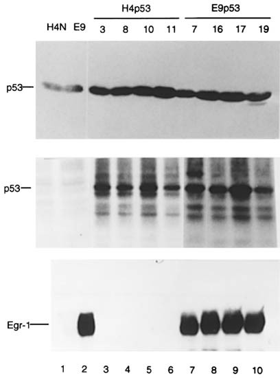
The human fibrosarcoma cell line, HT1080, clone H4, was used to determine if the transformation suppressive functions of p53 and Egr-1 have the same underlying mechanism. This cell line
expresses only mutant p53 and no detectable Egr-1. H4 clones stably expressing Egr-1 are less transformed in proportion to the level of Egr-1 expressed, acting through the induction of the
TGFβ1 gene. Here, H4 cells and the highest Egr-1 expressing clone were transfected with a vector expressing normal human p53 to derive stable clones expressing p53. The expression of p53 in
H4 cells inhibited transformed growth and reduced tumorigenicity. The effect of co-expression of both p53 and Egr-1 was additive, producing cell lines with 30% of normal growth rate and
sevenfold reduced tumorigenicity compared with control lines. These results indicated that each factor may act independently by different pathways, although each additively increased the
level of p21WAF1 cell cycle inhibitor. However, exposure of the H4-derived cells to UV-C irradiation produced contrasting effects. Cell cycle analyses showed that the presence of p53 was
associated with loss of the G1 and S cells to apoptosis after irradiation. In contrast, the expression of Egr-1 increased entry into S/G2 phase of the cell cycle with little apoptosis via a
mechanism involving elevated FAK and low caspase activities. Apoptosis was observed only in the cell lines that expressed no Egr-1, especially those expressing wt-p53, and was preceded by
high caspase activity. In summary, Egr-1 suppressed transformation and counteracted apoptosis by the coordinated activation of TGFβ1, FN, p21 and FAK, leading to enhanced cell attachment and
reduced caspase activity. In the doubly expressing cell line, the survival effect of Egr-1 was dominant over the apoptotic effect of p53.
We thank our colleagues at the Burnham Institute, S Frisch, A Marti, JC Reed, I Tamm and X Zhang, for gifts of antibodies, for advice and for critical comments on the manuscript. We are
indebted to B Vogelstein and J Trill for plasmids, and to JT Parsons for antibodies to PYK-2. We are grateful for support from the Public Health Services, grant CA 67888 (ED Adamson), CA
63783 and CA76173, (D Mercola) from the National Cancer Institute.
Ian de Belle and Ruo-Pan Huang: I de Belle and R-P Huang made equal contributions to this work
La Jolla Cancer Research Center, The Burnham Institute, 10901, N Torrey Pines Road, La Jolla, CA 92037, California, USA
Molecular Medicine, Northwest Hospital, 120 Northgate Plaza, Suite 230, Seattle, WA 98125, Washington, USA
Sidney Kimmel Cancer Center, 10835 Altman Row, San Diego, CA 92121, California, USA
Center for Molecular Genetics, University of California at San Diego, CA 92093, California, USA
Anyone you share the following link with will be able to read this content: