Multiple extradural arachnoid cysts at the spinal cord and cauda equina levels in the young
- Select a language for the TTS:
- UK English Female
- UK English Male
- US English Female
- US English Male
- Australian Female
- Australian Male
- Language selected: (auto detect) - EN
Play all audios:

ABSTRACT STUDY DESIGN: A case report of multiple extradural arachnoid cysts at the spinal cord and cauda equina levels in the young. OBJECTIVE: To report an exceedingly rare case of multiple
extradural arachnoid cysts at the spinal cord and cauda equina levels in the young. SETTING: Department of Orthopaedic Surgery, Tokai, Japan. CASE REPORT: An 11-year-old boy was diagnosed
with multiple extradural arachnoid cysts at the spinal cord and cauda equina levels extending from the T5 to L5 vertebrae and surgery was performed. At 2 years after surgery, no recurrence
was observed and muscle weakness of the lower extremities and sensory disturbance improved. CONCLUSION: Excision of only the arachnoid cysts at the spinal cord level led to a favorable
outcome. SIMILAR CONTENT BEING VIEWED BY OTHERS A CASE OF NEURENTERIC CYST OF SPINE MIMICKING AN ARACHNOID CYST Article 14 March 2022 SURGICAL TREATMENT FOR RECURRENT THORACIC VENTRAL
INTRADURAL ARACHNOID CYST SECONDARY TO TUBERCULOUS MENINGITIS: A CASE REPORT Article 23 May 2024 IMAGE CHARACTERISTICS OF RETAINED MEDULLARY CORD IN SECONDARY NEURULATION ARREST: AN
OBSERVATIONAL STUDY Article Open access 27 November 2024 INTRODUCTION Spinal arachnoid cyst is a relatively rare disease that accounts for approximately 1% of primary spinal cord tumors;
however, the widespread use of magnetic resonance imaging (MRI) has been associated with an increasing number of reported cases in recent years. Here we report a case of multiple extradural
arachnoid cysts at the spinal cord and cauda equina levels in the young. CASE REPORT The patient was an 11-year-old boy who had a chief complaint of gait disturbance without a significant
family history. He was born prematurely and weighed 1155 g at birth without gross external congenital anomaly; there have been no known growth abnormalities. At 1 month before being referred
to our institution, he had a contusion of the buttocks when falling from stairs at school and a diagnosis of coccygeal fracture was made, which was treated conservatively at another clinic.
About a week later, gait disturbance emerged and he was referred to our institution for suspected spinal epidural hematoma. Examination showed hyperesthesia at the level of T8–T10 and
hypesthesia at the level of T10 and below, together with weakness of the lower extremities MRC grade 3–4 on manual muscle testing. Increased deep tendon reflexes of the lower extremities and
ankle clonus were observed; Babinski reflex was present. Plain radiographs on admission revealed a deformity suggesting apophyseal separation in the L1 vertebra and spina bifida in the L5
vertebra. An anteroposterior radiograph of the thoracic and lumbar vertebrae showed increased interpedicular distance, while a lateral radiograph revealed increased anteroposterior diameter
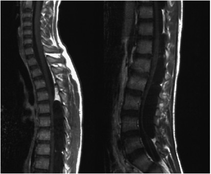
of the spinal canal. MRI disclosed multiple cystic mass densities, which were isointense to cerebrospinal fluid, extending from the upper margin of T5 to the upper margin of L5 on the dorsal
aspect of the spinal cord and cauda equina (Figure 1). Myelography and subsequent computed tomography (CT) showed leakage of contrast medium into the cysts disclosed by MRI (Figure 2).
These findings led to a diagnosis of multiple extradural arachnoid cysts at the spinal cord and cauda equina levels extending from the T5 to L5 vertebrae, and surgery was performed.
Laminectomy was performed from the T5 to T9 vertebrae and from the T11 to L1 vertebrae. Arachnoid cysts were identified: (1) from the upper margin of T5 to the upper margin of T8, (2) from
the upper margin of T8 to the upper margin of T10, (3) from the lower margin of T10 to the lower margin of T12, (4) from the lower margin of T12 to the upper margin of L2, and (5) the upper
margin of L2 and below. The communications between the above-mentioned cysts and the dura mater were found as follows: (1) in the left T7 nerve root, (2) in the left T8 nerve root, (3) from
T11 to T12 vertebrae on the left, and (4) from T12 to L1 vertebrae on the right (Figure 3). The communications (1) and (2) were ligated and the corresponding arachnoid cysts were excised.
The arachnoid cysts (3) and (4) were excised, the dura mater sutured, and the cysts removed. The arachnoid cyst located at the level of L2 and below was left untreated. Histopathological
examination of the cyst wall showed layered collagenous fibers and epithelial tissue with flat lining cells, confirming arachnoid cysts (Figure 4). As of this writing, 2 years after surgery,
no recurrence was observed and muscle weakness of the lower extremities and sensory disturbance improved. However, scoliosis with a Cobb angle of 16° was found between the T7 and L1
vertebrae. Between the T2 and L1 vertebrae, backward curvature with a preoperative Cobb angle of 28° changed to 62° postoperatively, without significant effects on his growth. DISCUSSION
Spinal arachnoid cyst was first reported by Spiller in 1903.1 Although spinal arachnoid cyst is a benign tumor accounting for approximately 1% of overall spinal cord tumors,2 incidence of
the disease has been frequently reported in recent years because of the development of MRI. Most of the reported cases are of solitary cysts occurring commonly at the thoracic level in
individuals in a wide range of ages from 10 to 69 years. Extradural arachnoid cysts may be caused by congenital or acquired factors, including inflammation, trauma, and iatrogenic ones,
although most cases are believed to be congenital in origin. In all cases, the disease results from sliding of the arachnoid from defective or fragile dura mater.2 The arachnoid may be
enlarged by the ball-valve mechanism of the nerve root or change in spinal pressure associated with pulse, breathing, and coughing.3, 4 Clinical course should be closely monitored in
asymptomatic individuals, while surgical treatment is indicated in patients with significant neurologic findings; complete closure of the communication is essential for complete cure. Dorsal
spinal arachnoid cysts may be associated with fragility of spinal posterior supporting tissues, requiring consideration of spinal reconstruction. There have been only five reported cases of
multiple arachnoid cysts, including this case, regardless of intradural or extradural ones (Table 1).2, 5, 6, 7 Multiple arachnoid cysts occurred in a narrow age group from 9 to 12 years of
age, and their affected sites varied widely from the thoracic to sacral vertebrae. In spinal development, union of the vertebral arch occurs by approximately 7 years of age, and the cricoid
cartilage develops all along the vertebrae at approximately 10 years of age, followed by longitudinal growth of the vertebrae.8, 9, 10 From this fact, we speculate that, multiple arachnoid
cysts predominantly occur in this age group because the longitudinal growth of the vertebrae is involved in the onset of the disease, together with extensively and ventrally compressed
spinal cord. Regarding treatment, three of the four patients with multiple arachnoid cysts previously described in Cases 2–5 were surgically treated with laminoplasty. In Case 3, as with our
case, only excision of arachnoid cysts at the spinal cord level resulted in a favorable outcome. In all cases, kyphosis occurred postoperatively. In Case 4, recurrence was observed in the
arachnoid cyst with unclosed communication and the cyst with a shunt procedure, requiring additional surgery. In the present case, which involved extensive lesions in the young, the
arachnoid cysts at the spinal cord level were excised with their communications closed, and the arachnoid cyst at the cauda equina level was left untreated. Marked thinning of the vertebral
arch and externally extending arachnoid cysts made laminoplasty difficult; therefore, laminectomy was performed for excision of the arachnoid cysts. Although the patient had an uneventful
postoperative growth with improvement in neurological symptoms, residual scoliosis and kyphosis will require close follow-up. CONCLUSIONS We report a case of multiple extradural arachnoid
cysts at the spinal cord and cauda equina levels in the young, which was treated with excision of only the arachnoid cysts at the spinal cord level, leading to a favorable outcome.
REFERENCES * Spiller WG et al. A case of intradural spinal cyst with operation and recovery. _Trans Coll Physicians Philadelphia_ 1903; 25: 1–18. Google Scholar * Obara K et al. Multiple
arachnoid cysts in thoracic spine; a case report. _J Orthop Surg_ 2000; 51: 545–547. Google Scholar * McCrum C et al. Spinal extradural arachnoid pouches. _J Neurosurg_ 1982; 57: 849–852.
Article CAS Google Scholar * Rohrer DC et al. Intraspinal extradural meningeal cyst demonstrating ball-valve mecanism of formation. _J Neurosurg_ 1993; 78: 122–125. Article CAS Google
Scholar * Andrew WD et al. Spinal arachnoid cysts in children. _Radiology_ 1978; 126: 423–429. Article Google Scholar * Otake S et al. Multiple spinal arachnoid cyst (T4–L2); a case
report. _Kanto J Orthop Trauma_ 1995; 26: 88–91. Google Scholar * Lynn MM et al. Multiple extradural arachnoid cysts as a cause of spinal cord compression in a child. _J Neurosurg_ 1999;
91: 116–120. Google Scholar * Wesley WP . Development of the spine. In: Harry NH _et al_ (eds). _The Spine_. 3rd edn. WB Saunders: Philadelphia 1992, pp 3–33. Google Scholar * Mark B .
Normal spinal anatomy: normal sagittal plane alignment. In: Keith HB _et al_ (eds) _Spinal Surgery_. 2nd edn. Lippincott-Raven: Philadelphia 1997, pp 185–191. Google Scholar * Louis R .
_Development of the Spine. Surgery of the Spine_. Springer-Verlag: Berlin 1983, pp 4–34. Book Google Scholar Download references AUTHOR INFORMATION AUTHORS AND AFFILIATIONS * Department of
Orthopaedic Surgery, Surgical Science, Tokai University School of Medicine, Bohseidai, Isehara, Kanagawa, Japan T Takagaki, T Nomura, E Toh, M Watanabe & J Mochida Authors * T Takagaki
View author publications You can also search for this author inPubMed Google Scholar * T Nomura View author publications You can also search for this author inPubMed Google Scholar * E Toh
View author publications You can also search for this author inPubMed Google Scholar * M Watanabe View author publications You can also search for this author inPubMed Google Scholar * J
Mochida View author publications You can also search for this author inPubMed Google Scholar RIGHTS AND PERMISSIONS Reprints and permissions ABOUT THIS ARTICLE CITE THIS ARTICLE Takagaki,
T., Nomura, T., Toh, E. _et al._ Multiple extradural arachnoid cysts at the spinal cord and cauda equina levels in the young. _Spinal Cord_ 44, 59–62 (2006).
https://doi.org/10.1038/sj.sc.3101799 Download citation * Published: 05 July 2005 * Issue Date: 01 January 2006 * DOI: https://doi.org/10.1038/sj.sc.3101799 SHARE THIS ARTICLE Anyone you
share the following link with will be able to read this content: Get shareable link Sorry, a shareable link is not currently available for this article. Copy to clipboard Provided by the
Springer Nature SharedIt content-sharing initiative KEYWORDS * arachnoid cyst * extradural tumor * spinal cord tumor * cauda equina tumor