Dissection of mechanical force in living cells by super-resolved traction force microscopy
- Select a language for the TTS:
- UK English Female
- UK English Male
- US English Female
- US English Male
- Australian Female
- Australian Male
- Language selected: (auto detect) - EN
Play all audios:

ABSTRACT Cells continuously exert or respond to mechanical force. Measurement of these nanoscale forces is a major challenge in cell biology; yet such measurement is essential to the
understanding of cell regulation and function. Current methods for examining mechanical force generation either necessitate dedicated equipment or limit themselves to coarse-grained force
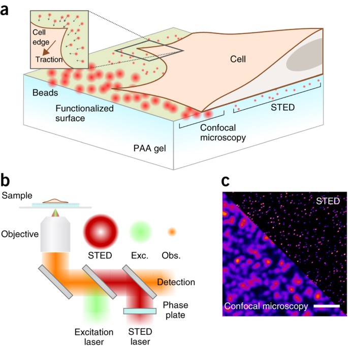
measurements on the micron scale. In this protocol, we describe stimulated emission depletion traction force microscopy—STED-TFM (STFM), which allows higher sampling of the forces generated
by the cell than conventional TFM, leading to a twofold increase in spatial resolution (of up to 500 nm). The procedure involves the preparation of functionalized polyacrylamide gels loaded
with fluorescent beads, as well as the acquisition of STED images and their analysis. We illustrate the approach using the example of HeLa cells expressing paxillin-EGFP to visualize focal
adhesions. Our protocol uses widely available laser-scanning confocal microscopes equipped with a conventional STED laser, open-source software and common molecular biology techniques. The
entire STFM experiment preparation, data acquisition and analysis require 2–3 d and could be completed by someone with minimal experience in molecular biology or biophysics. Access through
your institution Buy or subscribe This is a preview of subscription content, access via your institution ACCESS OPTIONS Access through your institution Access Nature and 54 other Nature
Portfolio journals Get Nature+, our best-value online-access subscription $29.99 / 30 days cancel any time Learn more Subscribe to this journal Receive 12 print issues and online access
$259.00 per year only $21.58 per issue Learn more Buy this article * Purchase on SpringerLink * Instant access to full article PDF Buy now Prices may be subject to local taxes which are
calculated during checkout ADDITIONAL ACCESS OPTIONS: * Log in * Learn about institutional subscriptions * Read our FAQs * Contact customer support SIMILAR CONTENT BEING VIEWED BY OTHERS
TWO-DIMENSIONAL TIRF-SIM–TRACTION FORCE MICROSCOPY (2D TIRF-SIM-TFM) Article Open access 12 April 2021 HIGH-RESOLUTION ASSESSMENT OF MULTIDIMENSIONAL CELLULAR MECHANICS USING LABEL-FREE
REFRACTIVE-INDEX TRACTION FORCE MICROSCOPY Article Open access 20 January 2024 ASTIGMATIC TRACTION FORCE MICROSCOPY (ATFM) Article Open access 12 April 2021 REFERENCES * Moeendarbary, E.
& Harris, A.R. Cell mechanics: principles, practices, and prospects. _Wiley Interdiscip. Rev. Syst. Biol. Med._ 6, 371–388 (2014). Article PubMed PubMed Central Google Scholar *
Capitanio, M. & Pavone, F.S. Interrogating biology with force: single molecule high-resolution measurements with optical tweezers. _Biophys. J._ 105, 1293–1303 (2013). Article CAS
PubMed PubMed Central Google Scholar * Bao, G. & Suresh, S. Cell and molecular mechanics of biological materials. _Nat. Mater._ 2, 715–725 (2003). Article CAS PubMed Google Scholar
* Janmey, P.A. & Miller, R.T. Mechanisms of mechanical signaling in development and disease. _J. Cell Sci._ 124, 9–18 (2011). Article CAS PubMed Google Scholar * Valero, C.,
Javierre, E., García-Aznar, J.M. & Gómez-Benito, M.J. A cell-regulatory mechanism involving feedback between contraction and tissue formation guides wound healing progression. _PLoS One_
9, e92774 (2014). Article PubMed PubMed Central CAS Google Scholar * Sasaki, N. in _Viscoelasticity - From Theory to Biological Applications_ 99–122 (InTech, 2012).
http://dx.doi.org/10.5772/49979. * Moeendarbary, E. et al. The cytoplasm of living cells behaves as a poroelastic material. _Nat. Mater._ 12, 253–261 (2013). Article CAS PubMed PubMed
Central Google Scholar * Fritzsche, M., Erlenkämper, C., Moeendarbary, E., Charras, G. & Kruse, K. Actin kinetics shapes cortical network structure and mechanics. _Sci. Adv._ 2, 1–13
(2016). Article CAS Google Scholar * Geiger, B., Spatz, J.P. & Bershadsky, A.D. Environmental sensing through focal adhesions. _Nat. Rev. Mol. Cell Biol._ 10, 21–33 (2009). Article
CAS PubMed Google Scholar * Sabass, B., Gardel, M.L., Waterman, C.M. & Schwarz, U.S. High resolution traction force microscopy based on experimental and computational advances.
_Biophys. J._ 94, 207–220 (2008). Article CAS PubMed Google Scholar * Colin-York, H. et al. Super-resolved traction force microscopy (STFM). _Nano Lett._ 16, 2633–2638 (2016). Article
CAS PubMed PubMed Central Google Scholar * Wilson, K. et al. Mechanisms of leading edge protrusion in interstitial migration. _Nat. Commun._ 4, 2896 (2013). Article PubMed CAS Google
Scholar * Dembo, M. & Wang, Y.-L. Stresses at the cell-to-substrate interface during locomotion of fibroblasts. _Biophys. J._ 76, 2307–2316 (1999). Article CAS PubMed PubMed Central
Google Scholar * Butler, J.P., Tolić-Nørrelykke, I.M., Fabry, B. & Fredberg, J.J. Traction fields, moments, and strain energy that cells exert on their surroundings. _Am. J. Physiol.
Cell Physiol._ 282, C595–C605 (2002). Article CAS PubMed Google Scholar * Judokusumo, E., Tabdanov, E., Kumari, S., Dustin, M.L. & Kam, L.C. Mechanosensing in T lymphocyte
activation. _Biophys. J._ 102, L5–L7 (2012). Article CAS PubMed PubMed Central Google Scholar * Fischer, R.S., Myers, K.A., Gardel, M.L. & Waterman, C.M. Stiffness-controlled
three-dimensional extracellular matrices for high-resolution imaging of cell behavior. _Nat. Protoc._ 7, 2056–2066 (2012). Article CAS PubMed Google Scholar * Damljanovic, V., Lagerholm,
B.C. & Jacobson, K. Bulk and micropatterned conjugation of extracellular matrix proteins to characterized polyacrylamide substrates for cell mechanotransduction assays. _Biotechniques_
39, 847–851 (2005). Article CAS PubMed Google Scholar * Gould, H.J. & Sutton, B.J. IgE in allergy and asthma today. _Nat. Rev. Immunol._ 8, 205–217 (2008). Article CAS PubMed
Google Scholar * Fritzsche, M. & Charras, G. Dissecting protein reaction dynamics in living cells by fluorescence recovery after photobleaching. _Nat. Protoc._ 10, 660–680 (2015).
Article CAS PubMed Google Scholar * Danuser, G., Allard, J. & Mogilner, A. Mathematical modeling of eukaryotic cell migration: insights beyond experiments. _Annu. Rev. Cell Dev.
Biol._ 29, 501–528 (2013). Article CAS PubMed PubMed Central Google Scholar * Lewalle, A. et al. A phenomenological density-scaling approach to lamellipodial actin dynamics. _Interface
Focus_ 4, 20140006 (2014). Article PubMed PubMed Central Google Scholar * Stachowiak, M.R. et al. Mechanism of cytokinetic contractile ring constriction in fission yeast. _Dev. Cell_ 29,
547–561 (2014). Article CAS PubMed PubMed Central Google Scholar * Pollard, T.D. Mechanics of cytokinesis in eukaryotes. _Curr. Opin. Cell Biol._ 22, 50–56 (2010). Article CAS PubMed
Google Scholar * Coward, J., Germain, R.N. & Altan-Bonnet, G. Perspectives for computer modeling in the study of T cell activation. _Cold Spring Harb. Perspect. Biol._ 2, a005538
(2010). Article PubMed PubMed Central CAS Google Scholar * Worth, A.J.J. et al. Disease-associated missense mutations in the EVH1 domain disrupt intrinsic WASp function causing
dysregulated actin dynamics and impaired dendritic cell migration. _Blood_ 121, 72–84 (2013). Article CAS PubMed PubMed Central Google Scholar * Stout, D.A. et al. Mean deformation
metrics for quantifying 3D cell–matrix interactions without requiring information about matrix material properties. _Proc. Natl. Acad. Sci. USA_ 113, 2898–2903 (2016). Article CAS PubMed
PubMed Central Google Scholar * Polacheck, W.J. & Chen, C.S. Measuring cell-generated forces: a guide to the available tools. _Nat. Methods_ 13, 415–423 (2016). Article CAS PubMed
PubMed Central Google Scholar * del Álamo, J.C. et al. Three-dimensional quantification of cellular traction forces and mechanosensing of thin substrata by Fourier traction force
microscopy. _PLoS One_ http://dx.doi.org/10.1371/journal.pone.0069850 (2013). * Thomas, D.G. et al. Non-muscle myosin IIB is critical for nuclear translocation during 3D invasion. _J. Cell
Biol._ 210, 583–594 (2015). Article CAS PubMed PubMed Central Google Scholar * Hell, S.W. & Wichmann, J. Breaking the diffraction resolution limit by stimulated emission:
stimulated-emission-depletion fluorescence microscopy. _Opt. Lett._ 19, 780–782 (1994). Article CAS PubMed Google Scholar * Gould, T.J., Kromann, E.B., Burke, D., Booth, M.J. &
Bewersdorf, J. Auto-aligning stimulated emission depletion microscope using adaptive optics. _Opt. Lett._ 38, 1860–1862 (2013). Article CAS PubMed PubMed Central Google Scholar *
Grashoff, C. et al. Measuring mechanical tension across vinculin reveals regulation of focal adhesion dynamics. _Nature_ 466, 263–266 (2010). Article CAS PubMed PubMed Central Google
Scholar * Ghassemi, S. et al. Cells test substrate rigidity by local contractions on submicrometer pillars. _Proc. Natl. Acad. Sci. USA_ 109, 5328–5333 (2012). Article CAS PubMed PubMed
Central Google Scholar * Vogel, V. & Sheetz, M. Local force and geometry sensing regulate cell functions. _Nat. Rev. Mol. Cell Biol._ 7, 265–275 (2006). Article CAS PubMed Google
Scholar * Blakely, B.L. et al. A DNA-based molecular probe for optically reporting cellular traction forces. _Nat. Methods_ 11, 1229–1232 (2014). Article CAS PubMed PubMed Central
Google Scholar * Stabley, D.R., Jurchenko, C., Marshall, S.S. & Salaita, K.S. Visualizing mechanical tension across membrane receptors with a fluorescent sensor. _Nat. Methods_ 9, 64–67
(2012). Article CAS Google Scholar * Liu, Y., Yehl, K., Narui, Y. & Salaita, K. Tension sensing nanoparticles for mechano-imaging at the living/nonliving interface. _J. Am. Chem.
Soc._ 135, 5320–5323 (2013). Article CAS PubMed PubMed Central Google Scholar * Wang, X. & Ha, T. Defining single molecular forces required to activate integrin and notch signaling.
_Science_ 340, 991–994 (2013). Article CAS PubMed PubMed Central Google Scholar * Morimatsu, M., Mekhdjian, A.H., Chang, A.C., Tan, S.J. & Dunn, A.R. Visualizing the interior
architecture of focal adhesions with high-resolution traction. _Nano Lett._ 15, 2220–2228 (2015). Article CAS PubMed PubMed Central Google Scholar * Müller, D.J. & Dufrêne, Y.F.
Atomic force microscopy as a multifunctional molecular toolbox in nanobiotechnology. _Nat. Nanotechnol._ 3, 261–269 (2008). Article PubMed CAS Google Scholar * Harris, A.R. &
Charras, G.T. Experimental validation of atomic force microscopy-based cell elasticity measurements. _Nanotechnology_ 22, 345102 (2011). Article PubMed CAS Google Scholar * Hu, K.H.
& Butte, M.J. T cell activation requires force generation. _J. Cell Biol._ 213, 535–542 (2016). Article CAS PubMed PubMed Central Google Scholar * Whited, A.M. & Park, P.S.-H.
Atomic force microscopy: a multifaceted tool to study membrane proteins and their interactions with ligands. _Biochim. Biophys. Acta._ 1838, 56–68 (2014). Article CAS PubMed Google
Scholar * Alcaraz, J. et al. Microrheology of human lung epithelial cells measured by atomic force microscopy. _Biophys. J._ 84, 2071–2079 (2003). Article CAS PubMed PubMed Central
Google Scholar * Darling, E.M., Topel, M., Zauscher, S., Vail, T.P. & Guilak, F. Viscoelastic properties of human mesenchymally-derived stem cells and primary osteoblasts, chondrocytes,
and adipocytes. _J. Biomech._ 41, 454–464 (2008). Article PubMed Google Scholar * Moreno-Flores, S., Benitez, R., Vivanco, M.d. & Toca-Herrera, J.L. Stress relaxation and creep on
living cells with the atomic force microscope: a means to calculate elastic moduli and viscosities of cell components. _Nanotechnology_ 21, 445101 (2010). Article PubMed CAS Google
Scholar * Guck, J. et al. Optical deformability as an inherent cell marker for testing malignant transformation and metastatic competence. _Biophys. J._ 88, 3689–3698 (2005). Article CAS
PubMed PubMed Central Google Scholar * Guck, J. et al. The optical stretcher: a novel laser tool to micromanipulate cells. _Biophys. J._ 81, 767–784 (2001). Article CAS PubMed PubMed
Central Google Scholar * Hochmuth, F.M., Shao, J.Y., Dai, J. & Sheetz, M.P. Deformation and flow of membrane into tethers extracted from neuronal growth cones. _Biophys. J._ 70,
358–369 (1996). Article CAS PubMed PubMed Central Google Scholar * Raucher, D. & Sheetz, M.P. Characteristics of a membrane reservoir buffering membrane tension. _Biophys. J._ 77,
1992–2002 (1999). Article CAS PubMed PubMed Central Google Scholar * Pontes, B. et al. Membrane elastic properties and cell function. _PLoS One_ 8, e67708 (2013). Article CAS PubMed
PubMed Central Google Scholar * Hochmuth, R.M. Micropipette aspiration of living cells. _J. Biomech._ 33, 15–22 (2000). Article CAS PubMed Google Scholar * Tseng, Y., Kole, T.P. &
Wirtz, D. Micromechanical mapping of live cells by multiple-particle-tracking microrheology. _Biophys. J._ 83, 3162–3176 (2002). Article CAS PubMed PubMed Central Google Scholar *
Scarcelli, G. et al. Noncontact three-dimensional mapping of intracellular hydromechanical properties by Brillouin microscopy. _Nat. Methods_ 12, 1132–1134 (2015). Article CAS PubMed
PubMed Central Google Scholar * Discher, D.E., Janmey, P. & Wang, Y.-L. Tissue cells feel and respond to the stiffness of their substrate. _Science_ 310, 1139–1143 (2005). Article CAS
PubMed Google Scholar * Tse, J.R., Engler, A.J., Tse, J.R. & Engler, A.J. Preparation of hydrogel substrates with tunable mechanical properties. _Curr. Protoc. Cell Biol._ CHAPTER
10, Unit 10.16 (2010). * Wen, J.H. et al. Interplay of matrix stiffness and protein tethering in stem cell differentiation. _Nat. Mater._ 13, 979–987 (2014). Article CAS PubMed PubMed
Central Google Scholar * Knoll, S.G., Ali, M.Y. & Saif, M.T. A novel method for localizing reporter fluorescent beads near the cell culture surface for traction force microscopy. _J.
Vis. Exp._ (91), 51873 (2014). * Clausen, M.P. et al. Pathways to optical STED microscopy. _NanoBioImaging_ 1, 1–12 (2013). Google Scholar * Vicidomini, G. et al. Sharper low-power STED
nanoscopy by time gating. _Nat. Methods_ 8, 571–573 (2011). Article CAS PubMed Google Scholar * Martiel, J.L. et al. Measurement of cell traction forces with ImageJ. _Methods Cell Biol._
125, 269–287 (2015). Article CAS PubMed Google Scholar * Schwarz, U.S. & Soiné, J.R.D. Traction force microscopy on soft elastic substrates: a guide to recent computational
advances. _Biochim. Biophys. Acta._ 1853, 3095–3104 (2015). Article CAS PubMed Google Scholar * Han, S.J., Oak, Y., Groisman, A. & Danuser, G. Traction microscopy to identify force
modulation in subresolution adhesions. _Nat. Methods_ 12, 653–656 (2015). Article CAS PubMed PubMed Central Google Scholar * Schwarz, U.S. et al. Calculation of forces at focal
adhesions from elastic substrate data: the effect of localized force and the need for regularization. _Biophys. J._ 83, 1380–1394 (2002). Article CAS PubMed PubMed Central Google Scholar
Download references ACKNOWLEDGEMENTS We gratefully acknowledge support from the Wolfson Imaging Centre (C. Lagerholm and E. Garcia) and funding from the Medical Research Council (MRC;
grant no. MC_UU_12010/unit programme G0902418 and grant no. MC_UU_12025), the MRC/Biotechnology and Biological Sciences Research Council (BBSRC)/Engineering and Physical Sciences Research
Council (EPSRC; grant no. MR/K01577X/1), the Wellcome Trust (grant ref. 104924/14/Z/14) and the Wolfson Foundation. We thank E. Sezgin for kindly reading the manuscript. AUTHOR INFORMATION
AUTHORS AND AFFILIATIONS * MRC Human Immunology Unit, Weatherall Institute of Molecular Medicine, University of Oxford, Oxford, UK Huw Colin-York, Christian Eggeling & Marco Fritzsche *
Kennedy Institute for Rheumatology, University of Oxford, Oxford, UK Marco Fritzsche Authors * Huw Colin-York View author publications You can also search for this author inPubMed Google
Scholar * Christian Eggeling View author publications You can also search for this author inPubMed Google Scholar * Marco Fritzsche View author publications You can also search for this
author inPubMed Google Scholar CONTRIBUTIONS H.C.-Y. conducted the experiments. H.C.-Y., M.F. and C.E. wrote the manuscript. CORRESPONDING AUTHOR Correspondence to Marco Fritzsche. ETHICS
DECLARATIONS COMPETING INTERESTS The authors declare no competing financial interests. RIGHTS AND PERMISSIONS Reprints and permissions ABOUT THIS ARTICLE CITE THIS ARTICLE Colin-York, H.,
Eggeling, C. & Fritzsche, M. Dissection of mechanical force in living cells by super-resolved traction force microscopy. _Nat Protoc_ 12, 783–796 (2017).
https://doi.org/10.1038/nprot.2017.009 Download citation * Published: 16 March 2017 * Issue Date: April 2017 * DOI: https://doi.org/10.1038/nprot.2017.009 SHARE THIS ARTICLE Anyone you share
the following link with will be able to read this content: Get shareable link Sorry, a shareable link is not currently available for this article. Copy to clipboard Provided by the Springer
Nature SharedIt content-sharing initiative
