Advances in bladder tissue engineering
- Select a language for the TTS:
- UK English Female
- UK English Male
- US English Female
- US English Male
- Australian Female
- Australian Male
- Language selected: (auto detect) - EN
Play all audios:

Jack, G. S. _ et al_. Urinary bladder smooth muscle engineered from adipose stem cells and a three dimensional synthetic composite. _Biomaterials_ 30, 3259–3270 (2009). Long Heise, R. _ et
al_. Generating elastin-rich SIS-based smooth muscle constructs utilizing exogenous growth factors and cyclic mechanical stimulation. _Tissue Eng. Part A_ doi:10.1089/ten.TEA.2009.0044 Two
new studies highlight the ongoing progress in tissue engineering for bladder augmentation surgery, by showcasing the potential for novel cell types and culture conditions to enhance the
growth and function of tissue-engineered grafts. The first study, published in _Biomaterials_, demonstrates the possibility of using adipose tissue as a source of stem cells for bladder
tissue engineering. Adipose-derived stem cells can be directed to differentiate into a range of lineages including functioning smooth muscle _in vitro_, and they offer the advantage of being
both accessible and abundant. The investigators obtained adipose stem cells from liposuction procedures and differentiated them into smooth muscle cells using inductive media. These cells
were seeded onto synthetic bladder scaffolds, which were used to augment the bladders of adult female athymic rats that had undergone partial cystectomy to remove 50% of the urinary bladder.
Other animals received augmentation with unseeded bladder scaffolds or suture closure with no augmentation. By 12 weeks after surgery, implanted rats had organized urothelium covering the
entire lumenal surface of the scaffold, as well as evidence of differentiated and organized smooth muscle structures. The amount of smooth muscle was greatest in the rats with cell-seeded
scaffolds, and these animals also demonstrated less fibrosis and collagen deposition than the animals with acellular implants. After organ harvesting, the cell-seeded tissue demonstrated a
weak contractile response to chemical stimulation. The authors conclude that adipose stem cells represent an attractive nonembryonic option for tissue engineering in the lower urinary tract.
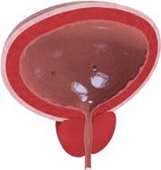
Credit: © Dreamstime The second study, from _Tissue Engineering Part A_, illustrates the importance of culture conditions in tissue engineering applications. Rather than use a synthetic
scaffold, the investigators focused on cell-seeded small intestinal submucosa (SIS). “In our previous work, we observed that while adult stem cells changed the mechanical properties of the
SIS, they did not penetrate into the tissue,” says Michael Sacks, corresponding author on the paper. “We thought that the best way to promote cellular penetration into the SIS was to use
growth factors that were important in development of the bladder.” In fact, addition of the growth factors VEGF and FGF-2 resulted in significant penetration of rat bladder smooth muscle
cells into SIS specimens. The researchers also studied the effect of mechanical stimulation, which is important to the normal development of the bladder, on the bioengineered tissue. Their
previous work had shown that _in vitro_ stretching of rat bladders at non-physiologic frequency promoted profound changes in the extracellular matrix, particularly in elastin content. “We
used this scheme on our engineered tissues once the cells had penetrated the matrix,” says Sacks. “We found that mechanical stimulation produced large amounts of elastin, which is important
in bladder compliance.” Thus, control of elastin deposition by _in vitro_ culture conditions and mechanical stimulation could be used to improve functional compliance in tissue engineered
bladder wall. Authors * Nick Groves-Kirkby View author publications You can also search for this author inPubMed Google Scholar RIGHTS AND PERMISSIONS Reprints and permissions ABOUT THIS
ARTICLE CITE THIS ARTICLE Groves-Kirkby, N. Advances in bladder tissue engineering. _Nat Rev Urol_ 6, 466 (2009). https://doi.org/10.1038/nrurol.2009.155 Download citation * Issue Date:
September 2009 * DOI: https://doi.org/10.1038/nrurol.2009.155 SHARE THIS ARTICLE Anyone you share the following link with will be able to read this content: Get shareable link Sorry, a
shareable link is not currently available for this article. Copy to clipboard Provided by the Springer Nature SharedIt content-sharing initiative