Door to the cell for covid-19 opened, leading way to therapies
- Select a language for the TTS:
- UK English Female
- UK English Male
- US English Female
- US English Male
- Australian Female
- Australian Male
- Language selected: (auto detect) - EN
Play all audios:

A VERY RECENT STUDY BY LAN ET AL.1PUBLISHED IN_NATURE_DETERMINED THE CRYSTAL STRUCTURE OF THE SEVERE ACUTE RESPIRATORY SYNDROME CORONAVIRUS (SARS-COV)-2 RECEPTOR-BINDING DOMAIN (RBD) BOUND
TO ANGIOTENSIN-CONVERTING ENZYME 2 (ACE2). THE STRUCTURE REVEALS THE MECHANISM OF SARS-COV-2 RBD RECOGNITION BY ITS RECEPTOR ACE2, WHICH IS HIGHLY CONSERVED IN ACE2 RECOGNITION OF SARS-COV
RBD. THE STUDY PROVIDES STRUCTURAL INFORMATION ON DEVELOPING SMALL MOLECULES TARGETING SARS-COV-2 RBD/ACE2 AND IMPLIES THE EXISTENCE OF OTHER MECHANISMS THAN RECEPTOR BINDING FOR THE
MARKEDLY DIFFERENT INFECTION ACTIVITY OF THE TWO EVOLUTIONARILY CLOSE VIRUSES. The outbreak of a novel and highly pathogenic coronavirus (SARS-CoV-2) has presented a serious global public
health emergency of coronavirus disease 2019. As of 11 May 2020, more than 4 million cases have been confirmed with the infection, leading to nearly 279,000 deaths in 214 countries
(https://www.who.int), and the coronavirus continues to spread quickly all over the world. Currently, effective vaccines or antiviral drugs for SARS-CoV-2 are unavailable. The newly
identified SARS-CoV-2 belongs to β-coronavirus, which also includes Middle East respiratory syndrome coronavirus (MERS-CoV) and SARS-CoV. The spike glycoprotein of coronaviruses acts as an
important determinant of their virulence activity by interacting with a receptor on the surface of host cells.2 The interaction between the spike glycoprotein and its receptor can serve as a
target for therapeutic interventions to treat diseases caused by coronaviruses. ACE2 has been identified as a functional receptor of SARS-CoV.3 More recently, ACE2 was also shown to be a
receptor of SARS-CoV-2.4 The ectodomain of the spike protein contains a receptor-binding unit S1 and a membrane-fusion unit S2. Interaction of the RBD from the S1 unit with ACE2 leads to
fusion of S2 with the host cell and viral membranes,2 thus mediating entry of coronavirus into host cells. To elucidate the mechanism of SARS-CoV-2 RBD and ACE2 interaction, Lan et al.1
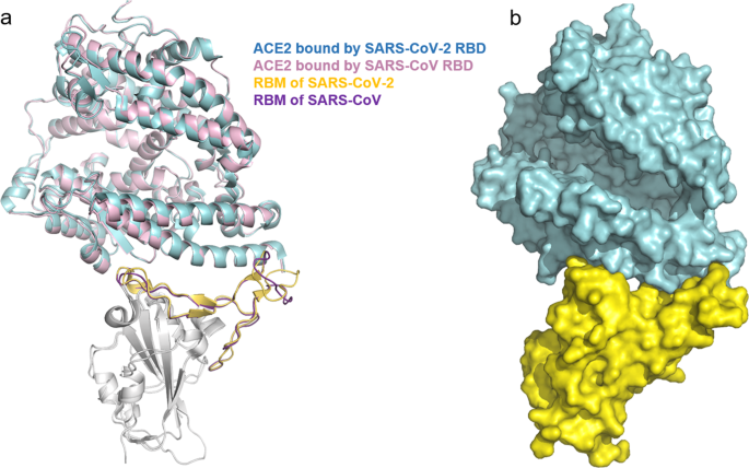
determined the complex structure of the two proteins at 2.45 Å resolution by X-ray crystallography. The final structural model contains residues of Thr333-Gly526 of the SARS-CoV-2 RBD, and
residues of Ser19-Asp615 of the ACE2 N-terminal peptidase domain (Fig. 1). The receptor-binding motif (RBM) at one side of SARS-CoV-2 RBD forms a concave for interaction with ACE2. The
overall structure of the SARS-CoV-2 RBD is highly similar to that of the SARS-CoV RBD.2 This is not surprising given 72% sequence identity of the two RBDs. However, remarkable conformational
differences occur to the loop from the distal end of the RBM that faces toward the solvent region. Structural comparison showed that the ACE2-bound SARS-CoV-2 RBD is nearly identical with
that from the free SARS-CoV-2 spike protein, indicating that ACE2 binding induces no notable conformational changes in SARS-CoV-2 RBD. Specific recognition of ACE2 by SARS-CoV-2 involves two
β-sheets (β5 and β6) and three connecting loops of the RBM. Structural superimposition showed that SARS-CoV-2 RBD and SARS-CoV RBD employ a highly conserved mechanism for interaction with
ACE2, supporting a close evolutionary relationship between the two viruses. Both of the RBM-ACE2 interfaces feature a large network of hydrogen bonds, highlighting specific interaction
between the two proteins. Fourteen of the ACE2-interacting residues are shared by the two RBDs, of which 8 are identical and 5 are similar. The shared but non-conserved residue interacts
with the same set of amino acids of ACE2. The conserved interactions in the SARS-CoV-2 RBM/ACE2 and SARS-CoV RBM/ACE2 complexes suggests that the two RBDs can have a similar affinity with
ACE2. Indeed, quantification assays using surface plasmon resonance showed that SARS-CoV-2 RBD and SARS-CoV RBD bound to the ACE2 receptor with an affinity ~4.7 and ~31.0 nM, respectively.
One notable difference between two complex structures is that Lys417 of SARS-CoV-2 is located outside the RBM but forms salt-bridge interactions with Asp30 of ACE2. By comparison, the
equivalent position of SARS-CoV has a valine residue, which is unlikely to form the salt bridges seen in the SARS-CoV-2 RBD/ACE2 complex. This subtle structural difference was proposed to
contribute to the slightly higher affinity between SARS-CoV-2 RBD and ACE2. However, ~20-fold difference between SARS-CoV-2 (Kd of 14.7 nM for ACE2) and SARS-CoV (Kd of 325 nM for ACE2) in
binding affinity with ACE2 was observed by another group,4 probably due to different proteins used in the assays. The study by Lan et al.1 also provided an explanation for the observation
that none of the isolated SARS-CoV monoclonal antibodies are able to neutralize SARS-CoV-2. By mapping the epitope residues of SARS-CoV RBD onto the sequence of SARS-CoV-2 RBD, they found
that one third (7 over 21) epitope positions of the antibody 80R are altered in SARS-CoV-2 RBD. A similar observation was also made with the epitope positions of the antibody m396.
Altogether, the study by Lan et al.1 revealed the structural mechanism of ACE2 receptor recognition by SARS-CoV-2 RBD. The mechanism sheds light on pathogenesis of the highly pathogenic
virus and can serve as a template for developing intervention strategies targeting SARS-CoV-2 spike and receptor recognition. It is unexpected that SARS-CoV-2 and SARS-CoV employ a nearly
identical mechanism for interaction of the ACE2 receptor, given their striking differences in infection and transmission activity.5 The mechanism underlying the differences remains poorly
understood, but the small difference between the two viruses in affinity with ACE2 is less likely to determine their distinct infection and transmission activity. As proposed by the authors,
some unique SARS-CoV-2-encoded proteins might have an important role in this aspect. It also remains possible that SARS-CoV-2 has other receptors(s) than ACE2 for entry into the host cells.
Addressing this question warrants future studies and will be conducive to developing antiviral therapies. REFERENCES * Lan, J. et al. Structure of the SARS-CoV-2 spike receptor-binding
domain bound to the ACE2 receptor. _Nature_ https://doi.org/10.1038/s41586-020-2180-5 (2020). * Li, F. Structure, function, and evolution of coronavirus spike proteins. _Annu. Rev. Virol._3,
237–261 (2016). Article CAS Google Scholar * Li, W. et al. Angiotensin-converting enzyme 2 is afunctional receptor for the SARS coronavirus. _Nature_426, 450–454 (2003). Article CAS
Google Scholar * Wrapp, D. et al. Cryo-EM structure of the 2019-nCoV spike in the prefusion conformation. _Science_367, 1260–1263 (2020). Article CAS Google Scholar * Tian, X. et al.
Potent binding of 2019 novel coronavirus spike protein by a SARS coronavirus-specific human monoclonal antibody. _Emerg. Microbes Infect._9, 382–385 (2020). Article CAS Google Scholar
Download references AUTHOR INFORMATION AUTHORS AND AFFILIATIONS * Center for Life Sciences, School of Life Science and Technology, Harbin Institute of Technology, Harbin, 150080, China
Zhiwei Huang * School of Life Sciences, Tsinghua University, Beijing, 100084, China Jijie Chai Authors * Zhiwei Huang View author publications You can also search for this author inPubMed
Google Scholar * Jijie Chai View author publications You can also search for this author inPubMed Google Scholar CORRESPONDING AUTHORS Correspondence to Zhiwei Huang or Jijie Chai. ETHICS
DECLARATIONS COMPETING INTERESTS The authors declare no competing interests. RIGHTS AND PERMISSIONS OPEN ACCESS This article is licensed under a Creative Commons Attribution 4.0
International License, which permits use, sharing, adaptation, distribution and reproduction in any medium or format, as long as you give appropriate credit to the original author(s) and the
source, provide a link to the Creative Commons license, and indicate if changes were made. The images or other third party material in this article are included in the article’s Creative
Commons license, unless indicated otherwise in a credit line to the material. If material is not included in the article’s Creative Commons license and your intended use is not permitted by
statutory regulation or exceeds the permitted use, you will need to obtain permission directly from the copyright holder. To view a copy of this license, visit
http://creativecommons.org/licenses/by/4.0/. Reprints and permissions ABOUT THIS ARTICLE CITE THIS ARTICLE Huang, Z., Chai, J. Door to the cell for COVID-19 opened, leading way to therapies.
_Sig Transduct Target Ther_ 5, 104 (2020). https://doi.org/10.1038/s41392-020-00215-6 Download citation * Received: 12 May 2020 * Revised: 28 May 2020 * Accepted: 02 June 2020 * Published:
26 June 2020 * DOI: https://doi.org/10.1038/s41392-020-00215-6 SHARE THIS ARTICLE Anyone you share the following link with will be able to read this content: Get shareable link Sorry, a
shareable link is not currently available for this article. Copy to clipboard Provided by the Springer Nature SharedIt content-sharing initiative