Differential effects of purified low molecular weight Poly(I:C) in the maternal immune activation model depend on the laboratory environment
- Select a language for the TTS:
- UK English Female
- UK English Male
- US English Female
- US English Male
- Australian Female
- Australian Male
- Language selected: (auto detect) - EN
Play all audios:

The Poly (I:C) (polyriboinosinic-polyribocytidilic acid) paradigm of maternal immune activation (MIA) is most widely used as experimental model for the evaluation of the effects of
gestational infection on the brain and behavior of the progeny. We have previously reported significant batch-to-batch variability in the effects of Poly (I:C), purchased from the same
supplier (Sigma–Aldrich), on maternal and fetal immune responses and found these differences to be dependent on the relative amount of synthetic double-stranded RNA fragments in the high
versus low molecular weight (LMW) range contained in the compound. We here resorted to Poly (I:C) purified for LMW dsRNA fragments to establish a MIA paradigm with increased reproducibility
and enhanced standardization in an effort to refine the MIA paradigm and characterize its effect on offspring behavior. We found that the parallel application of LMW Poly (I:C) in two
different MIA-experienced laboratories (Vienna and Zurich) yielded differential outcomes in terms of maternal immune responses and behavioral phenotypes in the offspring generation. In both
experimental sites, administration of LMW Poly (I:C) induced a significant sickness response and cytokine induction in the pregnant dam and fetal brains, while the expected deficit in
sociability as one main behavioral outcome parameter in the MIA progeny, was only present in the Zurich, but not the Vienna cohort. We conclude that although using Poly (I:C) purified for a
defined molecular weight range reduces batch-to-batch variability, it does not make the MIA model more reliable and robust. The differential response in behavioral phenotypes of the MIA
offspring between the two laboratories illustrates the highly complex interaction between prenatal and postnatal milieus - including the laboratory environment - that determine offspring
phenotypic outcomes after MIA. Consequently, establishing a new MIA protocol or implementing the MIA model firstly under new or changed environmental conditions must include the assessment
of offspring behavior to ensure solid and reproducible experimental outcomes.
In the past decades, animal models of maternal immune activation (MIA) have allowed considerable progress in our understanding of the involvement of gestational infection in the modulation
of fetal brain development and in shaping neurobehavioral outcomes in the progeny [1,2,3]. Epidemiological findings have repeatedly implicated maternal infection during pregnancy in an
increased risk for offspring neurodevelopmental disorders, including schizophrenia, autism spectrum disorders, and depressive disorders [4,5,6]. The establishment of reliable rodent models
enabled determining the underlying molecular mechanisms and cellular pathways and to derive causality between immune stimulation of the maternal system and deviation of offspring
neurodevelopment and its behavioral repercussions [7,8,9,10,11,12,13]. One of the most widely used substances to experimentally induce activation of the maternal immune system and to study
its effect independently of the immunogenic agent itself is Poly (I:C) (polyriboinosinic-polyribocytidilic acid). Poly (I:C) is a dsRNA analog whose administration causes a
cytokine-associated viral-like acute phase response [1]. The Poly (I:C) mouse model has been most successfully used in a large number of research studies to determine the pathophysiological
principles of MIA and comprehensively delineate its impact on brain function and behavior of the first, second, and even third-generation descendants [7,8,9,10,11,12,13]. While the Poly
(I:C) model has proven to be a very useful and effective tool for MIA research, it has become clear that the effect of Poly (I:C) in the MIA model may differ significantly between
experiments. Recently we have experimentally investigated the observation of substantial variability in results obtained when using different batches of Poly(I:C) supplied by the same vendor
[14]. In a comparative study across laboratories, we found that the relative composition of dsRNA fragment sizes of low molecular weight (LMW, 100–200 nucleotides) and high molecular weight
(1000–6000 nucleotides) determined Poly (I:C) immunogenicity and its impact on the stability of pregnancy, maternal and fetal immune responses. We concluded that using a Poly (I:C) product
with dsRNA fragments purified for their molecular weight may reduce the variability of research outcomes between different MIA experiments. However, although the standardization of the
molecular weight range of the Poly (I:C) product in MIA studies has been recommended to reduce variability [15], the impact of a Poly (I:C) preparation with defined molecular weight RNA
fragments on offspring behavior in the MIA paradigm has remained unexplored. While previous studies [15] reported more pronounced cytokine response and sickness behavior with HMW (high
molecular weight) Poly (I:C) in rats, we decided to investigate LMW Poly (I:C) only as a refinement measure, since recent studies including mice [14] showed that abortion rates are
considerably lower using LMW Pol
y (I:C) compared to HMW Poly (I:C). Here, we set out to establish a MIA protocol using a LMW Poly (I:C) product in parallel in two laboratories with ample experience in behavioral
phenotyping this model. We tested two different Poly (I:C) dosages, validated maternal sickness, cytokine response in the maternal and fetal systems, and characterized the behavioral
consequences in adult female and male offspring [14, 15].
C57Bl6/N male and female mice (Charles River, Sulzfeld, Germany) 10–12 weeks were used for experimental breeding (see Table 1 for detailed information about animal numbers). Upon arrival
from the vendor, all mice were housed in groups of 2–5 in individually ventilated cages in a specific-pathogen-free (SPF) holding room, which was temperature and humidity-controlled (21 ±
3°, 50 ± 10%) with a 12 h: 12 h light-dark cycle (Lights on 08:00 AM–08:00 PM (Vienna) and 09:00 PM–09.00 AM (Zurich)). All animals had ad libitum access to food (Ssniff Spezialdiäten,
Germany (Vienna) and Kliba AG, Switzerland (Zurich)) and water throughout the entire study. All animal experiments were conducted in agreement with the EU Directive 2010/63/EU for animal
experiments and approved by the Austrian (License number: BMBWF- 2021-0.150.413) and Swiss authorities (License number: ZH124/2020). Reporting of the animal experiments is in accordance with
the ARRIVE guidelines. All efforts were made to minimize the number of animals used and their burden.
Experimental breedings began after two weeks of habituation to the respective mouse vivarium using a previously described timed mating procedure (Khan et al., 2014, Mueller et al., 2018,
respectively). In Vienna, animals were mated for one single night only, and pregnancies were confirmed post-mating days 7–10 by the evaluation of weight gain of the dams. In Zurich, dams
were mated for several consecutive nights, and a plug check was performed each morning. Once the plug check was positive, the dam was moved to a new cage, and the preceding night was
considered the night of conception. The day of the single housing (Vienna) or observation of a vaginal plug (Zurich) was referred to as gestational day (GD) 0.5. Pregnant dams were randomly
assigned to either Poly (I:C) treatment or treatment with endotoxin-free 0.9% NaCl (B. Braun, Switzerland) vehicle solution. For all procedures, LMW Poly (I:C) (cat.#: tlrl-picw) was
obtained from Invivogen (USA). The same lot of Poly (I:C) (Lot #PIW-41-05) was used throughout the study for both laboratories.
Poly (I:C) was administered through a single intraperitoneal (i.p.) injection on GD 12.5 at 10 mg/kg or 20 mg/kg using an injection volume of 10 ml/kg. Immediately after Poly (I:C) or
vehicle administration, dams were placed back into their home cages and left undisturbed until the assessment of the sickness behavior and/or the collection of maternal and fetal tissues.
Offspring of Poly (I:C)- or vehicle-treated mothers were weaned on postnatal day (PND) 21 and littermates of the same sex were maintained in groups of 2–5 animals per cage. For the
reinfection experiment, Female F1 offspring (LMW Poly (I:C) and vehicle) were injected with LMW Poly (I:C) or vehicle on GD 12.5 in the F1 generation resulting in four groups (PIC/PIC;
PIC/CON; CON/PIC; CON/CON) for the evaluation of the F2 generation. Standardized detailed information is provided in the Reporting Guidelines Checklist in Supplementary Table 1 for Zurich
and Supplementary Table 2 for Vienna.
To assess signs of sickness in response to Poly (I:C) all animals were scored two hours after injection using predefined criteria [16], including body position, ptosis (drooping eyelids),
piloerection, coat condition, reaction to change in environment (cage, assessed by manual interaction within the cage) and nest condition. In both laboratories, sickness scores were
determined by one experimenter each, who was blinded to the experimental conditions. For every mouse and criterion, a value from 0 to 3 was assigned, with 0 indicating no signs of sickness
and 3 indicating the highest degree.
3 h post-injection, pregnant mice were decapitated, and trunk blood was collected in EDTA-coated tubes. Blood was kept on ice for less than. 20 min before centrifugation (10.000 rpm, 10 min,
4 °C) to collect plasma, which was stored at −20 °C until further use. Fetal brain tissues were collected as described before [17, 18]. In brief, the abdominal cavity of the dam was exposed
to collect the uterus, which was placed into a petri dish filled with ice-cold PBS. Decidual tissue and yolk sac were then removed for individual fetuses, and the fetuses were further
dissected to obtain fetal brain tissue according to landmarks shown in (Supplementary Fig. 1). Isolated fetal brain tissue was placed in Eppendorf tubes, snap-frozen by immersion in dry ice
and stored at −80 °C. Whenever possible, fetal brain tissue from two fetuses per uterine horn was collected for each pregnant dam by selecting fetuses in the most caudal uterine position.
Fetal brains from each dam were pooled and incubated in ice-cold Roche complete lysis buffer (Complete™ Lysis-M, Sigma–Aldrich, Switzerland) for 20 min before lysing using a tissue lyser
(tissue Lyser II, Quiagen®) for 3 min at a frequency of 20/s. After centrifugation (12,000 rpm at 4 °C for 20 min) the supernatants were removed and frozen and stored at −80 °C until the
cytokine assays were performed.
Cytokine levels in maternal plasma and fetal brains were quantified using a Meso-Scale Discovery (MSD) V-plex electrochemiluminescence assay for mice as previously described (Mueller et al.,
2018, 2019; Notter et al., 2018). The panel included interleukin (IL) IL-1β, IL-6, IL-10, tumor necrosis factor (TNF)-α and interferon (IFN)-γ. V-plex 96-well plates coated with primary
antibodies directed against the targeted cytokines were treated with the corresponding detecting antibodies, which were pre-labeled with SULFO-TAGTM (MSD, USA). The plates were read using
the MESO QuickPlex SQ 120MM (MSD) imager and analyzed using MSD’s Discovery Workbench analyzer and software package. Plasma and fetal brain lysates were diluted 2-fold and were run in
duplicates according to the manufacturer’s instructions. The detection limits were 0.11 pg/ml for IL-1β, 0.61 pg/ml for IL-6, 0.94 pg/ml for IL-10, 0.13 pg/ml for TNF-α and 0.04 pg/ml for
IFN-γ.
Behavioral testing was initiated when offspring reached 8–12 weeks of age and included tests for sociability (Social interaction test), innate anxiety behavior and locomotor activity (Open
field test) and Fear conditioning (second Vienna cohort only). These tests are widely used in animal models of MIA [5, 16, 19] and extensively validated [17, 20, 21]. The order of testing
was always kept constant (1. Open field test; 2. Social interaction test; 3. Fear conditioning – where applicable) and a resting interval of at least 3 days was kept between tests.
A standard open field test was applied to assess basal locomotor activity and innate anxiety-like behavior. The testing arena (40 × 40 cm) was made of white Plexiglas and surrounded by walls
(35 cm in height). The testing room was maintained at fixed lighting conditions (30 lx) and a digital camera was mounted directly above the arena. Activity was monitored by a computational
tracking system (Activity Monitor, MedAssociates, USA) in Vienna, and Ethovision (Noldus Information Technology, Netherlands) in Zurich, respectively. In each case, the animals were gently
placed in the center of the arena and allowed to freely explore for the testing duration of 10 min. For the purpose of data collection, the arena was conceptually partitioned into two areas:
a center zone measuring 15 × 15 cm2 in the middle of the area and a peripheral zone occupying the remaining area. The dependent measures were the total distance moved (cm) in the entire
arena.
Social interaction was determined by analyzing the relative exploration time between an unfamiliar congenic mouse and an inanimate object using a protocol established before [12, 21]. The
test apparatus was made of Plexiglas and consisted of three identical arms (Vienna: 37 cm × 6 cm × 16 cm; Zurich: 50 cm × 9 cm × 10 cm; length × width × height). The three arms radiated from
a centrally located equilateral triangle spaced 120° from each other. Two out of the three arms contained a rectangular stranger cage. The third arm did not contain a cage and served as the
starting zone (see below). All animals were habituated to the test apparatus on the day before social interaction testing for 5 min. During the test phase on the following day, one stranger
cage contained an unfamiliar C57BL6/N mouse of the same sex (10–12 weeks of age), whereas the other stranger cage contained an inanimate dummy object. This inanimate dummy object consisted
of three black Lego bricks in Vienna and a black scrunchy in Zurich. The allocation of the unfamiliar live mouse and inanimate dummy object to the two stranger cages was counterbalanced
across experimental groups. To start a test trial, the test mouse was gently placed in the starting arm and allowed to explore freely for 5 min. A digital camera was mounted above the test
apparatus which captured and transmitted images to the EthoVision tracking system. Behavioral observations were made by an experimenter who was blinded to the experimental conditions, and
social interaction was defined as nose contact within a 5-cm interaction zone. For each animal, the relative time spent with the unfamiliar mouse was calculated by the formula [(time spent
with the mouse) / (time spent with the inanimate object + time spent with the mouse)] × 100.
In the Vienna cohort, a standard Pavlovian conditioning protocol was employed as previously reported [11]. Briefly, mice were handled by the experimenter on the two days directly preceding
the start of the FC paradigm. On trial days, mice were allowed to habituate for approximately 45 min in a holding room adjacent to the experimental room. The 12 min conditioning training
session consisted of the delivery of three pairings between an unconditioned stimulus (US: foot shock, 0.3 mA, 1 s) and a conditioned stimulus (CS: white noise, 75 dB, 30 s) with an
inter-trial interval of 180 s. Cued recall was tested 24 h after the training session in a test trial lasting 720 s in total, with the preCS phase being 360 s long while the CS was presented
for 180 s, followed by a postCS phase of 180. The response to the CS was evaluated as the freezing levels during the presentation of the CS (preCS-CS). Freezing behavior (percentage of time
spent freezing, defined as lack of movement for at least 0.5 s, or 15 frames at a sampling rate of 30 frames/s) was automatically recorded using the ‘NIR Video Fear Conditioning Contextual
Package for Mouse’ system and accompanying Video Freeze software (Med Associates, USA).
Statistical analysis was performed in the R statistical programming environment [22]. For comparing treatment effects on cytokine concentration in maternal plasma, two-way type 1 ANOVAs with
lab and treatment and the lab-by-treatment interaction were used. Separate tests were run for cohorts of dams receiving 10 and 20 mg/kg Poly (I:C). Behavioral measures and sickness scores
were compared with three-way or two-way type 1 ANOVAs with lab, treatment, and sex (not for sickness score) and all interaction terms as fixed factors. After finding a significant
interaction effect of lab and treatment for social interaction, post-hoc two-way ANOVAs with treatment and sex and the sex-by-treatment interaction were done for each lab. Additional mixed
effect models using the package lme4 [23] including dam as random factor showed that dam explained only a negligible proportion of the overall variance and its inclusion did not affect
outcomes for the main factors. We identified multivariate outliers for the behavioral measures using the package mvoutlier. Re-calculating ANOVA statistics after the removal of these
outliers did not change the outcomes for treatment, lab, and lab-by-treatment interaction, corroborating the robustness of the results. For investigating treatment effects in a new cohort of
dams receiving 20 mg/kg at the lab in Vienna, we used two-way type 1 ANOVAs with treatment, sex, and sex-by-treatment interaction as fixed factors. Re-calculating ANOVA statistics after the
removal of multivariate outliers did, again, not change the outcome for treatment effects. For analyzing treatment effects in the F2 generation after reinfection, we used three-way type 1
ANOVAs with the treatment of dams in F0 and F1, sex, and all interaction terms as fixed factors. Re-calculating ANOVA statistics after the removal of multivariate outliers did not change the
outcome for treatment effects. For comparing treatment effects on cytokine concentration in fetal brains, two-way type 1 ANOVAs with lab and treatment and the lab-by-treatment interaction
were used. Separate tests were run for cohorts of dams receiving 10 and 20 mg/kg. Assumptions for ANOVA were checked by visually inspecting QQ-plots and scale location plots using the
package lindia [24]. QQ-plots showed that residual distributions for cytokines in dam plasma and brain tissue were partly heavy-tailed but symmetrical. Residuals for social interaction were
slightly left-skewed and light-tailed, though given that the observed residuals fitted the prediction line well over a wide range, data transformation was not considered necessary.
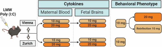
As a first step in the establishment of the LMW Poly (I:C) MIA model (Fig. 1) we set out to determine the immediate immune response of the pregnant female to the Poly (I:C) challenge at GD
12.5 (Fig. 2A). To this end we monitored sickness behavior of the dam and evaluated maternal plasma levels of a panel of cytokines previously characterized in our and others´ MIA studies (eg
[14, 25,26,27]). The selected cytokines comprised IL-1β, IL-6, IL-10, TNF-α, and IFN-γ, which have not only been found dysregulated in the MIA paradigm, but also form part of the
inflammatory profiles in the neurodevelopmental disorders reflected in the MIA model (eg [28,29,30,31]). Three groups (10 mg/kg or 20 mg/kg Poly (I:C) and vehicle controls) of animals were
included in both labs (Vienna and Zurich). We focused on comparisons of either dosage (10 mg/kg or 20 mg/kg) Poly (I:C) to controls (treatment effect), while also monitoring possible
differences between the Vienna and the Zurich sites (lab effect) and interactions between the above (lab x treatment interactions). For sickness behavior we found a significant treatment
effect for the 10 and 20 mg/kg dosage (F(1,33) = 33.758, p