Matrix stiffness changes affect astrocyte phenotype in an in vitro injury model
- Select a language for the TTS:
- UK English Female
- UK English Male
- US English Female
- US English Male
- Australian Female
- Australian Male
- Language selected: (auto detect) - EN
Play all audios:

Injury to the central nervous system (CNS) usually leads to the activation of astrocytes, followed by glial scar formation. The formation of glial scars from active astrocytes in vivo has
been found to be dependent on the cell microenvironment. However, how astrocytes respond to different microenvironmental cues during scar formation, such as changes in matrix stiffness,
remains elusive. In this work, we established an in vitro model to assess the responses of astrocytes to matrix stiffness changes that may be related to pathophysiology. The investigated
hydrogel backbones are composed of collagen type I and alginate. The stiffness of these hybrid hydrogels can be dynamically changed by association or dissociation of alginate chains through
adding crosslinkers of calcium chloride or a decrosslinker of sodium citrate, respectively. We found that astrocytes obtain different phenotypes when cultured in hydrogels of different
stiffnesses. The obtained phenotypes can be switched in situ when changing matrix stiffness in the presence of cells. Specifically, matrix stiffening reverts astrogliosis, whereas matrix
softening initiates astrocytic activation in 3D. Moreover, the effect of matrix stiffness on astrocytic activation is mediated by Yes-associated protein (YAP), where YAP inhibition enhances
the upregulation of GFAP and contributes to astrogliosis. To investigate the underlying mechanism of matrix stiffness-dependent GFAP expression, we also developed a mathematical model to
describe the time-dependent dynamics of biomolecules involved in the matrix stiffness mechanotransduction process of astrocytes. The modeling results further indicate that the effect of
matrix stiffness on cell fate and behavior may be related to changes in the cytoskeleton and subsequent activity of YAP. The results from this study will guide researchers to re-examine the
role of matrix stiffness in reactive astrogliosis in vivo and inspire the development of a novel therapeutic approach for controlling glial scar formation following injury, enabling axonal
regrowth and improving functional recovery by exploiting the benefits of mechanobiology studies.
Injury to the central nervous system (CNS) usually induces astrocytic reactivity, followed by glial scar formation1. For years, glial scarring has been thought to be a major obstacle to
successful axon regeneration2,3. However, recently accumulated evidence suggests a beneficial role for this scar tissue as part of the endogenous local immune regulation and repair
process1,4,5. At an early stage after injury, the glial scar protects damaged neural tissue by preventing an overwhelming inflammatory response and reestablishing the blood–brain
barrier6,7,8. On the other hand, at later stages, scarred tissue releases biochemical factors (e.g., chondroitin, heparan, dermatan, and keratan sulfate proteoglycans8), which may impair
axonal outgrowth, causing aberrant function or death of neurons6,7,8. As a result, it is crucial to regulate the formation of glial scars during treatment of neural injury.
Moreover, as the key effector cells in glial scar formation, astrocytes have been found to change their phenotype from naive astrocytes to reactive astrocytes and gradually into scar-forming
astrocytes, upregulating glial fibrillary acidic protein (GFAP)9,10,11 and vimentin12 and inflammatory proteins such as IL-1β13. Such astrocytic phenotype transformation has been found to
depend on the cell microenvironment in vivo14. For example, reactive astrocytes could revert in retrograde to naive astrocytes in a proper microenvironment. The phenotypic changes of
astrocytes are affected by multiple microenvironmental cues, one of which is matrix stiffness15,16,17,18. Glial scars in both the rat cortex and spinal cord are significantly softer than
those in healthy CNS tissues6. Several in vitro models have been developed to study the influences of matrix stiffness on astrocyte phenotypic changes15,16,17,18. For example, by culturing
astrocytes on substrates of different stiffnesses, it was found that a stiff substrate induces the activation of astrocytes15,16,17. However, the influences of dynamic changes in matrix
stiffness on astrocyte behaviors have not been reported, although matrix stiffness usually changes dynamically during glial scar formation. Recently, the phenotype of astrocytes plated on
reversible fibrous supramolecular hydrogel was found to be capable of switching reversibly, where the reversible hydrogel can undergo reversible formation and disappearance of
superstructures18. However, the relationship between astrocyte phenotypic switching and hydrogel stiffness changes remains elusive since the mechanical and structural changes of the hydrogel
are not decoupled. Moreover, the above studies were established in 2D culture14,15,16,18,19,20,21, which failed to recapitulate the native 3D cell microenvironment. Therefore, it is still
of great interest to study the effects of dynamic changes in matrix stiffness on astrocyte phenotypic switching in 3D culture.
In this work, we established an in vitro model to study the effects of dynamically changing matrix stiffness on astrocyte phenotypic switching in 3D, which is related to the glial
scar-forming process. The hybrid hydrogels were composed of collagen type I, a representative matrix under pathological conditions, and alginate, an inherently bioinert material that was
introduced to dynamically tune the mechanical properties of the hydrogel. The responses of astrocytes to different matrix stiffnesses were evaluated by characterizing the expression of GFAP
and interleukin 1 beta (IL-1β). The stiffness of the hybrid hydrogels was further tuned in situ to investigate the effects of matrix stiffening and softening on astrocyte phenotypic
switching. We found that astrocytes develop an activated phenotype in soft hydrogels but maintain an inactive phenotype in stiff hydrogels. Stiffening soft hydrogels reverts the activation
of astrocytes, whereas softening the stiff matrix induces astrocyte activation. Yes-associated protein (YAP) was found to be involved in matrix stiffness-mediated astrocyte activation. We
also developed a mathematical model to study the underlying mechanism of matrix stiffness-dependent GFAP expression, and the modeling results were consistent with the experimental results.
Based on the mathematical model, we further speculate that the effect of matrix stiffness on cell fate and behavior may be related to changes in the cytoskeleton and the subsequent activity
of YAP. These results may improve the understanding of astrocyte mechanobiology and inspire the development of novel therapeutic approaches for treating CNS injury.
Recent studies have proven that the stiffness of glial scars is significantly lower than that of healthy neural tissue, which is associated with increased concentrations of softer
extracellular matrix (ECM) components6 (e.g., CSPGs22 and GAGs23). The elastic stiffness of the cortex can change from ~1 kPa (healthy tissue) to nearly ~50 Pa (scar region)6. To mimic the
dynamic change in the native cell mechanical microenvironment, we fabricated a set of hybrid hydrogel systems composed of collagen type I and alginate with dynamically tunable stiffness.
Collagen type I, the major component of hybrid hydrogels, has been highlighted in many studies for its advantageous features in supporting cell adhesion, growth, and migration24,25,26,27.
Recent studies also proved that the proportion of collagen in the ECM significantly increases after brain injury, which contributes to the scar formation process6,8,14,18. Moreover, rat
primary astrocytes cultured in proper concentrations of collagen type I can maintain a relatively naive phenotype compared to those of those cultured in 2D28,29,30. The addition of alginate
supports cells in a round or spreading state with the same matrix stiffness due to its nonadhesive and nanoporous structures. The most attractive characteristic of alginate hydrogels is that
alginate can be crosslinked or decrosslinked reversibly to dynamically tune the mechanical properties of the hydrogel27,31. In this work, the stiffness of the hybrid hydrogels was
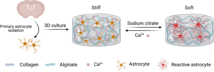
dynamically tuned by crosslinking or decrosslinking alginate with calcium chloride or sodium citrate, respectively (Fig. 1a). According to rheological measurements, the storage moduli of the
hybrid hydrogels were found to be tuned from ~42.7 ± 6.3 Pa (soft) to ~990.6 ± 189.3 Pa (stiff) (Fig. 1a), which strongly matches the native situations6. We measured the softened and
stiffened hydrogels with a rheometer at the same time (Supplementary Fig. 1c). We also applied a custom-designed magnetic tweezer to define the mechanical properties of the inner hydrogels
and found that the stiffness was consistent with the rheological results (Supplementary Fig. 1d). By analyzing the swelling performance of the hybrid hydrogels, we found that the swelling
ratio of the hybrid hydrogels increased with the addition of increasing concentrations of Ca2+ (Supplementary Fig. 1a). Moreover, we evaluated the internal microstructure of the hybrid
hydrogels by scanning electron microscopy (SEM) and found that alginate acts as a filler between collagen fibers (Supplementary Fig. 1b).
a Astrocytes were encapsulated into hybrid hydrogels of different stiffnesses. The mechanical properties of the hybrid hydrogel changed from ∼42.7 ± 6.3 Pa (soft) to ~990.6 ± 189.3 Pa
(stiff) when the concentration of Ca2+ increased from 0 to 5 mM. b The cell spreading area significantly increased with decreasing matrix stiffness. c Matrix stiffness influenced the shape
of astrocytes. d, e Immunofluorescence analysis indicated that astrocytes showed increased activity in a softer matrix with upregulated GFAP and IL-1β after 7 days of culture (red, IL-1β;
green, GFAP; blue, nucleus). Scale bar, 50 μm. f Relative protein levels of GFAP in astrocytes determined by Western blotting. n > 50 cells for (b–e).
To assess the biocompatibility of the hybrid hydrogels for culturing astrocytes in 3D, primary astrocytes were isolated from the cortex of neonatal rats, expanded in vitro, and encapsulated
into the hydrogels during gelation. The average viability of the encapsulated astrocytes was >90% after culture for 14 days (Supplementary Fig. 2a, b), indicating high biocompatibility of
the hybrid hydrogels. In addition, decreasing matrix stiffness resulted in a reduced proliferation rate of astrocytes (Supplementary Fig. 2c, d). Further, the proliferation rate of
astrocytes decreases during culture, consistent with previous studies32.
Several studies have shown that after brain injuries, naive astrocytes transform into reactive astrocytes and contribute to glial scar formation1,33,34,35,36. It has been reported that
matrix stiffness acts is an important factor in astrocytic activation; however, most previous studies on matrix stiffness regulating astrocytic activation were unidirectional and performed
in 2D culture14,15,16,18,19,20,21. To explore the influences of matrix stiffness on astrocyte activation in 3D, we encapsulated astrocytes into hybrid hydrogels of different stiffnesses
(i.e., stiff, moderate and soft), which were cultured for 7 days. From F-actin staining (Supplementary Fig. 3), we found that astrocytes cultured in the stiff matrix showed a round
morphology with a smaller spreading area, while astrocytes in the softer matrix showed characteristic hypertrophy of cell bodies with a much larger spreading area and a prominent stretched
morphology (Fig. 1b). In addition, the cell shape index of astrocytes dropped significantly with decreasing matrix stiffness (Fig. 1c). We further observed that astrocytes cultured in soft
hydrogels showed significantly upregulated expression of GFAP and IL-1β compared to those cultured in stiff hydrogels for 7 and even 14 days. (Fig. 1d, e, and Supplementary Figs. 4, 5). The
higher expression levels of GFAP in the soft matrix were further confirmed by Western blot analysis (Fig. 1f). Collectively, these results suggest that the phenotype and activation of
astrocytes in 3D can be regulated by matrix stiffness.
To confirm this result, we seeded astrocytes on stiff, moderate and soft matrices. We found that astrocytes seeded on the soft matrix showed a reactive phenotype with upregulated GFAP, while
astrocytes seeded on the stiff matrix restored the naive phenotype with rounded morphology and low expression of GFAP (Supplementary Fig. 6). The results proved that matrix stiffness does
have a strong influence on the activity of astrocytes under both 2D and 3D conditions. In particular, the soft matrix activates astrocytes from a naive phenotype to a reactive phenotype.
To exploit the effects of dynamically changing matrix stiffness on astrocyte phenotypic switching, we first cultured astrocytes in stiff and soft matrices for 7 days individually. Then, we
softened the stiff matrix in situ and stiffened the soft matrix. The astrocyte phenotype was evaluated after another 7 days of culture after dynamic softening or stiffening of the matrix. We
observed that astrocytes switch from a naive to a reactive phenotype after softening the stiff matrix by decrosslinking alginate with sodium citrate (Fig. 2) and from a reactive to a naive
phenotype after stiffening the soft matrix by crosslinking alginate with calcium chloride (Fig. 3). Specifically, astrocytes cultured in the stiff matrix maintained a naive phenotype with a
rounded morphology and low expression of astrocyte markers (GFAP and IL-1β) in the first 7 days of culture. After softening the stiff matrix, astrocytes switch to a typical reactive
phenotype with hypertrophy of cell bodies (Supplementary Fig. 7a), an increased spreading area (Fig. 2b) and significantly upregulated the expression of GFAP and IL-1β (Fig. 2d, e). In
addition, the higher expression levels of GFAP in the soft matrix were further confirmed by Western blot analysis (Fig. 2f), where the expression of GFAP in the softened matrix was ~1.5
times higher than that in the stiff matrix. In addition, we found that astrocytes reverted to a native phenotype after stiffening the soft matrix (Fig. 3 and Supplementary Fig. 7b). Taken
together, our results indicate that dynamic changes in matrix stiffness can switch the astrocyte phenotype in 3D; specifically, matrix softening induces astrocyte activation, while matrix
stiffening reverts the activation of astrocytes.
a Schematic depiction of matrix softening. b The cell spreading area significantly increased when the matrix was softened after 7 days of culture. c Matrix softening influenced the shape of
astrocytes. d, e Immunofluorescence analysis indicated that the activation of astrocytes increased when the matrix was softened after 7 days (red, IL-1β; green, GFAP; blue, nucleus). Scale
bar, 50 μm. f Relative protein levels of GFAP determined by Western blotting in astrocytes after 7 days and 14 days of culture.
a Schematic depiction of matrix stiffening. b The cell spreading area significantly decreased when the matrix was stiffened after 7 days of culture. c Matrix stiffening influenced the shape
of astrocytes. d, e Immunofluorescence analysis indicated that the activation of astrocytes decreased when the matrix was stiffened after 7 days (red, IL-1β; green, GFAP; blue, nucleus).
Scale bar, 50 μm. f Relative protein levels of GFAP determined by Western blotting in astrocytes after 7 days and 14 days of culture.
To explore the effect of chemical components, such as calcium chloride and sodium citrate, on astrocyte behavior, P1 astrocytes were seeded at a density of 1 × 10⁴ cells/cm2 on plastic
culture plates and treated individually with 10 mM calcium chloride and 10 mM sodium citrate for 30 min. Since astrocytes spread over the plate in 5 days, we compared the GFAP expression of
cells in the experimental groups after culture for 1, 3, and 5 days with the no treatment control group (Supplementary Fig. 8). We found that neither calcium chloride nor sodium citrate had
a significant influence on the phenotype of astrocytes, as reflected by GFAP levels (Supplementary Fig. 8b, c).
Since we added 10% (wt/vol) alginate and an equal volume of 10 mM calcium chloride into the soft hydrogel to stiffen the soft hydrogel, it was important to clarify whether the excess
alginate would cover the RGD domain in the collagen, which may prevent adhesion and extension of the cells. We seeded P1 astrocytes in hybrid hydrogels containing 5% (wt/vol) and 10%
(wt/vol) alginate (final concentrations of 5%, 10% (wt/vol) alginate, 1 mg/mL collagen, and 0 mM Ca2+). We first measured the stiffness of both hydrogels and found no significant difference
between the two hydrogels (Supplementary Fig. 9b). According to the analysis of GFAP expression, we found that excess uncrosslinked alginate did not prevent the reactivity of astrocytes, and
there was also no significant difference between the two groups (Supplementary Fig. 9a, c). Together, our data proved that the added chemicals and excess uncrosslinked alginate did not
affect the phenotype of astrocytes. It is only the stiffness of the substrate that regulates astrocyte phenotype in 3D.
Although matrix stiffness clearly regulates astrocyte phenotypic transformation18, the underlying mechanoregulatory pathways are still poorly understood. In the 3D hybrid hydrogel system, we
noticed that the enhanced activation of astrocytes in the soft matrix was always accompanied by an extended spreading area (Supplementary Figs. 3, 7). According to previous studies, matrix
stiffness affects cell fate and behavior by changing cytoskeletal tension37,38,39,40 and rapidly remodeling the cytoskeleton41. To test this possibility, we first cultured astrocytes on
stiff matrix for 3 days, treated astrocytes with a series of concentrations of blebbistain to inhibit cytoskeletal tension to different degrees and cultured the astrocytes until the 14th
day. Our data suggest that astrocytes treated with blebbistain exhibit an extended spreading area and filopodium-rich morphology along with significant upregulation of GFAP (Supplementary
Fig. 10). Moreover, the influence of blebbistain on astrocyte activation is dependent on both dose and time. Thus, the inhibition of cytoskeletal tension induces the activation of
astrocytes, suggesting that matrix stiffness might affect astrocyte activation by regulating cytoskeletal tension.
YAP42,43, a key regulator in multiple organ development44, is highly expressed in astrocytes in vivo. To test whether YAP activity is regulated by matrix stiffness, we characterized the
expression of YAP in astrocytes cultured in stiff and soft matrices. We found that astrocytes cultured in the stiff matrix displayed higher levels of YAP expression than those cultured in
the soft matrix after 7 days of culture. We also noticed that the nuclear localization of YAP was increased in astrocytes cultured in the stiff matrix (Fig. 4a–d). Then, we analyzed the
expression of YAP and GFAP in astrocytes cultured in matrix with dynamic stiffness changes. We found that astrocytes in the stiff matrix maintained a naive phenotype with low expression of
GFAP and rather high activity of YAP, while after matrix softening, naive astrocytes switched to reactive astrocytes with upregulated GFAP and decreased YAP expression (Fig. 4a). In
addition, the corresponding phenotypic transformation of astrocytes was also observed when stiffening the soft matrix (Fig. 4b). The results indicate that the expression of YAP is
sensitively regulated by matrix stiffness and its dynamic changes. They also indicate that YAP could be involved in the responses of astrocyte phenotype switching to dynamically changing
matrix stiffness.
a, b Immunofluorescence analysis suggested that increasing matrix stiffness reduced the expression of the YAP protein and upregulated the expression of the GFAP protein (red, YAP; green,
GFAP; blue, nucleus). Scale bar, 50 μm. c Quantification of the ratio of nuclear YAP to cytoplasmic YAP in astrocytes. d Quantification of the normalized total YAP levels. e
Immunofluorescence analysis indicated that astrocyte activation increased when astrocytes in a stiff matrix were transfected with siRNAs targeting YAP (blue, nucleus; green, GFAP). Scale
bar, 50 μm. f Quantification of GFAP levels when astrocytes in a stiff matrix were transfected with siRNAs targeting YAP.
To gain insight into the role of YAP in the responses of astrocytes to matrix stiffness, we transfected astrocytes cultured for 3 days with siRNAs targeting YAP (SiYAP) to inhibit the
expression of YAP. Four days later, we found that astrocytes transfected with SiYAP showed enhanced activation with upregulation of GFAP in the stiff matrix compared to the control group
(Fig. 4e, f). These results indicate that YAP might be a key factor in the response of astrocytes to matrix stiffness.
To investigate the underlying mechanism by which matrix stiffness affects GFAP expression, we also developed a mathematical model consisting of a series of ordinary differential equations
(ODEs) that describe the time-dependent dynamics of biomolecular interactions45,46. The modeling results showed that a stiff matrix inhibits the expression of GFAP through the activation of
YAP. We further compared the predicted normalized GFAP levels from our mathematical model (Fig. 5c) with the experimental results (Fig. 1d, e). We found that the modeling and experimental
results were consistent.
a The signaling pathway of the mathematical model. b The expression levels of YAP varied with time under different matrix stiffnesses (42.3, 307.6, and 990.6 Pa). c Simulated results for
GFAP levels are shown. As the matrix stiffness increases, GFAP expression decreases. d Sensitivity analysis of the model parameters in the study of how matrix stiffness affects GFAP
expression.
To evaluate the roles of intracellular signaling dynamics in matrix stiffness-mediated GFAP expression in astrocytes, we performed parameter sensitivity analysis using the developed
mathematical model (Fig. 5d). We divided the parameters into the following three categories according to the key biomolecules involved in mediating GFAP expression: (1) molecules associated
with the cell membrane; (2) molecules associated with the cytoskeleton; and (3) molecules associated with the nucleus. The sensitivity analysis of parameters showed that parameters related
to the cytoskeleton and nucleus have a greater impact on GFAP expression. Based on this, we speculate that matrix stiffness may induce changes in cytoskeletal tension, which may further
affect the activity of YAP and thus GFAP expression. This is consistent with the results reported in the literature20,37,38,39,40,41,42,47.
Reactive astrogliosis, which ultimately forms glial scars, was not thought to be reversible until an in vivo study revealed the microenvironment-dependent plasticity of astrocyte phenotypes,
where reactive astrocytes could revert in retrograde to naive astrocytes in an appropriate microenvironment14. We first proved the plasticity of astrocyte phenotypes in a 3D in vitro model
from a mechanobiological perspective. Our findings show that matrix stiffness is a vital microenvironmental cue that impacts astrocyte phenotypic transformation. We found that astrocytes
developed an activated phenotype in soft hydrogels but maintained an inactive phenotype in stiff hydrogels (Fig. 1d–g). Interestingly, stiffening of soft hydrogels reverted the activation of
astrocytes, whereas softening of the stiff matrix induced astrocyte activation (Figs. 2, 3). Thus, we linked the phenotypic transformation of astrocytes to dynamic changes in matrix
stiffness for the first time. Moreover, according to an in vivo study, glial scars in the rat cortex are significantly softer than healthy CNS tissues, as measured by atomic force
microscopy6. Our results therefore mimic this phenomenon and suggest that the softer components of the ECM (such as highly hydrated CSPGs) increase in abundance during scar formation, which
may induce astrocyte activation. Furthermore, this reactive astrogliosis could be reverted when the matrix environment is restored to the preinjury structure.
Collagen is produced when the CNS is injured48,49. We noted that during CNS scar formation, the stiffness of the CNS also changes. Both the changed collagen content and matrix stiffness
could contribute to astrocyte activation and scar formation. While collagen has been widely used as an ECM mimic for culturing diverse types of cells (including astrocytes), the effects of
matrix stiffness changes on astrocyte behavior in pathological conditions remain unclear. Therefore, we used collagen as a representative matrix in pathological conditions for culturing
astrocytes. Because it is inherently bioactive and easy to obtain, collagen has been applied as a brain engineering scaffold for diverse clinical indications. For instance, bioengineered
brain-like cortical tissue based on collagen type I gel and silk scaffolds promoted neuron anchoring and the formation of neural networks50. Collagen type I was used in coculturing
endothelial cells, pericytes, and astrocytes to model the blood–brain barrier in 3D51. Moreover, neural progenitors cultured in collagen type I gels showed significantly higher survival and
neuronal differentiation than those cultured on 2D gels52. Additionally, astrocytes seeded in the collagen type I system remained in a low reactive state, resembling their status in the
healthy CNS28,29. We acknowledge that simply using collagen is not sufficiently representative of the complex CNS matrix in pathological conditions. Hyaluronic acid and laminin could be
incorporated into collagen to better establish pathological relevance systems53,54,55,56. Further work is needed to investigate this.
In addition, to decouple the effects of matrix stiffness from collagen content, we used Ca2+-crosslinked alginate (an inherently bioinert material) to dynamically tune the mechanical
properties of the hydrogel without significantly changing the collagen content or structure. It is important to develop new methods and material systems to independently tune the mechanical
properties of hydrogels, avoiding the use of nonnative ECM components such as alginate.
Injury to the CNS activates naive astrocytes to reactive astrocytes. Recent evidence has indicated that reactive astrocytes can be further differentiated into A1 reactive astrocytes and A2
reactive astrocytes according to their functions57,58,59,60,61,62. The toxic A1 phenotype, which can be marked by complement component 3 (C3), induces neuronal death and shows a broader
relevance to neurodegenerative diseases57,58,59,60,62,63. In addition, neuroprotective A2 astrocytes, marked by S100A10, contribute to protecting neurons and repairing damage61,63. In this
study, we mainly focused on the process from of activation from naive astrocytes to reactive astrocytes. We actually did not aim to distinguish reactive astrocytes from the A1 phenotype to
the A2 phenotype. To characterize the ratio of type A1 or type A2 astrocytes to total cells, astrocytes enveloped in both stiff and soft hybrid hydrogels were subjected to colocalization of
C3 and S100A10 after culture for 1 and 7 days. We found that A2 reactive astrocytes were very rare in each group (almost none). After 7 days, the number of cells of the A1 phenotype
significantly increased in the soft hydrogel compared to the stiff hydrogel (Supplementary Fig. 11), which matched the results shown in Fig. 1d and Supplementary Fig. 4. In particular, rare
astrocytes seeded in stiff hydrogels were marked by C3 or S100A10 on day 1, which means that the astrocytes maintained a naive phenotype, consistent with the results in Fig. 1d and
Supplementary Fig. 4. Our results also proved that astrocytes were naive at the beginning of the experiments.
In addition to the characteristic marker GFAP, we also chose the IL-1β protein to reflect the inflammatory response of astrocytes. IL-1β is widely regarded as the top level of the cascade of
neuroinflammatory mediators13,64 and is associated with astrocyte activation65,66,67. In this work, we found that astrocytes cultured in soft hydrogels showed significantly upregulated
expression of IL-1β and GFAP (Fig. 1d). A recent study reported that A1 reactive astrocytes could increase their expression of proinflammatory cytokines, such as IL-1β68. We also detected
that most of the reactive astrocytes were A1 type (Supplementary Fig. 11), which is consistent with our results (Fig. 1d and Supplementary Figs. 4, 5).
Matrix stiffness affects cell fate and behavior by changing cytoskeletal tension and rapidly remodeling the cytoskeleton. In our study, we observed that a stiff matrix prevents the spreading
of astrocytes along with low expression of GFAP. The inhibition of cytoskeletal tension induced the activation of astrocytes cultured on stiff matrix, exhibiting an extended spreading area,
filopodium-rich morphology, and significant upregulation of GFAP (Supplementary Fig. 10). This implies that cytoskeletal tension might be involved in the regulation of the astrocyte
phenotype by matrix stiffness.
YAP, a key regulator in multiple organ development, has been reported to be associated with negative control of reactive astrogliosis. Our results indicate that the activity of YAP is
regulated by matrix stiffness and its dynamic changes (Fig. 4a–d). Further exploration proved that the decrease in YAP expression enhances reactive astrogliosis with upregulation of GFAP
(Fig. 4e, f), which indicates that YAP might be a critical factor for the astrocyte response to dynamically changing matrix stiffness. Combining the sensitivity analysis with the
mathematical model, we speculate that matrix stiffness may induce changes in cytoskeletal tension, which may further affect the activity of YAP and thus the astrocytic phenotype (through
GFAP expression). Thus, these findings suggest the potential mechanism by which matrix stiffness regulates astrocyte phenotypic transformation.
Matrix stiffness has been proven to be a major mechanical cue in modulating cell behaviors, for example, governing stem cell fate69. Cells cense dynamic mechanical changes via deformation
and reorganization of the microtubule cytoskeleton, and the degree of matrix stiffening determines cell fate69. Our results support this point, and we found that astrocyte fate can be
controlled by matrix stiffness in a 3D microenvironment. In addition to matrix stiffness, dynamic architectural features of hydrogel networks could also be involved in regulating the
astrocytic phenotype18. Here, we decouple the mechanical and structural changes of hydrogels and clarify the effect of the mechanical response (dynamically changed matrix stiffness) of
hydrogels on cell phenotypic transformation. Our hydrogel system imitates the modulus (~40–1000 Pa) of both healthy and injured brain tissues in vivo and provides a physiologically relevant
3D microenvironment for astrocytes, which allows us to assess the impact of dynamically changing stiffness on astrocytic phenotypic transformation. Moreover, the dynamic hydrogel system
would contribute to further explorations of the engineering of dynamic cellular microenvironments.
In this study, we established an in vitro model to assess the responses of astrocytes to matrix stiffness changes that may be related to pathophysiology. We found that astrocytes exhibit
different phenotypes when cultured in hydrogels of different stiffnesses. More importantly, the adopted phenotypes can be switched in situ when matrix stiffness is changed in the presence of
cells, i.e., the cells can adapt to the in situ changes in matrix stiffness by switching their phenotypes. Specifically, we showed that matrix stiffening can revert astrogliosis, whereas
matrix softening induces astrocyte activation in 3D. We also revealed that YAP is involved in matrix stiffness-regulated astrocyte activation, where the inhibition of YAP induces the
upregulation of GFAP and contributes to astrogliosis. We further developed a mathematical model to study the underlying mechanism by which matrix stiffness affects GFAP expression, and the
modeling results were consistent with the experimental results. The findings from this study may enhance the understanding of the role of mechanical cues in reactive astrogliosis and inspire
the development of a potential therapeutic approach for controlling glial scar formation following injury from the mechanobiological perspective.
To synthesize hybrid hydrogels with tunable stiffness, 20% (wt/vol) alginate and 4 mg/mL collagen type I were first mixed before adding CaSO4 suspension (0, 8, and 20 mM, respectively) and
cell culture medium to reach a final concentration of 5% (wt/vol) alginate, 1 mg/mL collagen, and 0/2/5 mM Ca2+. The mixture was then incubated for 45 min at 37 °C to allow crosslink
formation. Following the literature27, hydrogel softening was achieved by chelating Ca2+ with 100 mM sodium citrate for 30 min at 37 °C to dissolve alginate, followed by washing with cell
culture medium. To stiffen the hydrogels, 10% (wt/vol) alginate was added on the surface of soft hydrogels. After 45 min of infiltration at 37 °C, the excess alginate was removed, and an
equal volume of 10 mM calcium chloride was added to crosslink alginate infiltrated into the collagen.
To test the rheology of the hybrid hydrogels, samples were prepared as disks with a diameter of 8 mm and a thickness of 2 mm. Before rheological testing, the hybrid hydrogels were soaked in
PBS for 24 h. At the beginning of the test, the plate slowly descended until reaching the normal force of ~20 mN. Frequency tests were applied with a strain of 0.5% and a frequency ranging
from 0.1 to 10 rad/s. The storage modulus and loss modulus were calculated from the average of the modulus between frequencies of 0.25 and 2.5 rad/s, where the modulus maintained a balanced
value.
A simple magnetic tweezer device was used to determine the modulus of the material. Briefly, an electromagnet with a sharp-tipped nickel alloy core generated a strong magnetic field
gradient, thus creating an attractive force on the bead that points toward the tip. The magnetic force applied on the bead was calibrated according to the speed of the beads in a viscous
liquid as a function of the tip-to-bead distance. Bead positions were recorded by a CCD camera and tracked using a grayscale binarized center-of-mass algorithm in MATLAB. The strain (ε) of
the materials was defined as the ratio of the displacement of the beads (x) to the diameter of the beads (2 R), and the stress (σ) was defined as the ratio of the applied magnetic force (F)
to the maximum cross-sectional area of the beads:
After being fixed with 2.5% glutaraldehyde for 2 h, all samples were dehydrated by exposure to a graded series of ethanol-t-butanol mixtures (ethanol/t-butanol = 70/30, 50/50, 30/70, 20/80,
and 10/90 for 10 min each) before being treated with pure t-butanol. After being dried by a freeze dryer, the samples were coated with gold. Imaging was performed by employing a MAIA3 LMH
field emission SEM at 15 kV accelerating voltage.
After freeze-drying, the dry weight (Wd) of the samples was determined by using an electronic precision balance. After that, the samples were placed in PBS to swell for 24 h at room
temperature. The surface moisture of the samples was removed by filter papers before weighing the wet weight (Ww) of the samples. The swelling ratio of the samples was calculated as (Ww −
Wd)/Wd × 100%.
All Sprague-Dawley (SD) rats used in the present study were obtained from the Laboratory Animal Center of Xi’an Jiaotong University Health Science Center, China. All procedures and ethics
guidelines were approved by the Committee for Experimental Animal Use and Care of Xi’an Jiaotong University Health Science Center, China. Astrocytes were collected from the cortex of
neonatal rats (1 day old). Briefly, the cortical tissues of neonatal rats were dissociated with trypsin, and cells were plated in a 75 cm2 flask (Thermo Fisher Scientific, USA) in Dulbecco’s
modified Eagle’s medium with F-12 nutrient mixture (DMEM/F-12; HyClone, USA) containing 10% fetal bovine serum (FBS; Gibco, USA), 1% penicillin-streptomycin (Pen-Strep; Gibco, USA) at 37 °C
and 5% CO2. After 24 h, the culture medium was changed twice per week. Cultures were shaken at 300 rpm/min for 12–18 h to remove other types of neuronal cells after 10 days. All experiments
were performed using astrocytes from the first passage (P1). P1 cells were seeded at a density of 1 × 10⁶ cells/mL in combined hydrogels and cultured in DMEM/F-12 medium containing 10% FBS
and 1% Pen-Strep for 7 or 14 days.
For cell viability experiments, samples were incubated in 1 mL of PBS containing 0.5 μL of Calcein-AM (Life Technologies, USA) and 2 μL of EthD-1 (Life Technologies, USA) for 15 min at 37 °C
and 5% CO2, followed by three washes with PBS. Imaging was performed with an OLYMPUS IX81 inverted phase contrast fluorescence microscope. Live and dead cells were counted by using a cell
counter plug-in for ImageJ.
Samples were fixed in 4% paraformaldehyde for 20 min and washed in PBS three times for 5 min before penetrating samples with 0.5% Triton X-100 (Sigma, USA) for 15 min and washed in PBS three
times for 5 min. Samples were then incubated in PBS containing 10% normal goat serum (Thermo Fisher Scientific, USA) and 5% bovine serum albumin (BSA; MP Biomedicals, USA) for 1 h. Samples
were incubated in primary antibodies prepared in proper dilutions in PBS containing 1% BSA overnight at 4 °C. After washing with PBS, the samples were incubated with appropriate Alexa 488 or
Alexa 594 secondary antibodies (1:400, Thermo Fisher Scientific, USA). To visualize F-actin, samples were stained with Alexa 488-labeled phalloidin for 1 h. The following primary antibodies
were used for immunofluorescence: rabbit anti-IL-1β (inflammatory marker, 1:100; Abcam, ab9722, USA), mouse anti-GFAP (1:400; Cell Signaling Technology, 3670, USA), rabbit anti-Ki67
(proliferation marker, 1:400; Cell Signaling Technology, 9129, USA), rabbit anti-S100A10 (1:500; Bioss, bs-8503R, China), mouse anti-C3 (1:200; Santa Cruz, sc-8399, USA), and rabbit anti-YAP
(1:100; Cell Signaling Technology, 14074, USA). After labeling nuclei with DAPI (1 μg/mL; Sigma, D9542, USA) for 10 min, fluorescence images were taken with an Olympus FV3000 laser scanning
confocal microscope.
Based on the F-actin immunofluorescence staining of samples, the cell shape index was analyzed with Fuji software (ImageJ); the higher the cell shape index was, the closer to a circle the
cell was.
C = 4_pi_A/P^2–12.57_A/P^2, where C is the circularity, A is the area, and P is the perimeter.
Confocal images were taken with distinct z-stacks. For all the samples, the distance between two z-stacks was set to the same value. For the quantitative analysis of GFAP, YAP and nuclear
localization of YAP, the fluorescence intensity of all images was set to the same parameter. The normalized protein levels of the samples were analyzed by Fuji software with the Image
plugin.
Astrocytes were cultured in hydrogels for 3 days, and then YAP knockdown was performed in astrocytes by using a small interfering RNA (siRNA). Control cells were transfected with control
siRNA (5′-UUCUCCGAACGUGUCACGUTT). siRNA transfections were performed with Lipofectamine™ 2000 (Life Technologies, USA) in Opti-MEM I reduced serum medium (Invitrogen, USA) according to the
manufacturer’s instructions. The target sequence of the siRNA was 5′-GGUCAGAGAUACUUCUUAATT for rat YAP.
For Western blot analysis, protein extracts were obtained from samples cultured after 1, 7, or 14 days. First, the samples were treated with 100 mM sodium citrate for 30 min to dissolve
alginate, followed by washing with PBS. Then, 5 mg/mL type IV collagenase was added to dissolve collagen. The total protein extracts were separated by SDS-polyacrylamide gel electrophoresis
and electrotransferred to polyvinylidene difluoride membranes (Millipore, USA). The membranes were blocked with TBST containing 5% BSA and incubated with primary antibodies overnight at 4
°C. The following antibodies were used: rabbit anti-GAPDH (1:1000; Cell Signaling Technology, 2118, USA), GFAP (1:1000; Abcam, USA), and YAP (1:1000; Cell Signaling Technology, 14074, USA).
Then, the membranes were washed with TBST five times and incubated with the corresponding secondary antibodies (1:5000; Cell Signaling Technology, USA) for 2 h at room temperature. After
being washed with TBST five times, protein signals were detected by using a chemiluminescence system (ChemiScope 3300 mini, China). Densitometry analysis was performed with ImageJ (National
Institutes of Health, USA). The analyses were performed in three independent experiments with three replicates per condition.
Statistical analysis was conducted by using the OriginPro software package (Origin Pro 2016; Origin Lab, Northampton, USA). Statistics are presented as the mean ± standard deviation for all
quantitative data. One-way analysis of variance (ANOVA) was used for two-group comparisons along with a t-test (*p