High-speed imaging of glutamate release with genetically encoded sensors
- Select a language for the TTS:
- UK English Female
- UK English Male
- US English Female
- US English Male
- Australian Female
- Australian Male
- Language selected: (auto detect) - EN
Play all audios:

ABSTRACT The strength of an excitatory synapse depends on its ability to release glutamate and on the density of postsynaptic receptors. Genetically encoded glutamate indicators (GEGIs)
allow eavesdropping on synaptic transmission at the level of cleft glutamate to investigate properties of the release machinery in detail. Based on the sensor iGluSnFR, we recently developed
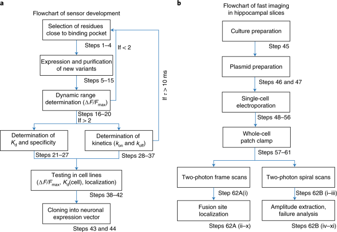
accelerated versions of GEGIs that allow investigation of synaptic release during 100-Hz trains. Here, we describe the detailed procedures for design and characterization of fast iGluSnFR
variants in vitro, transfection of pyramidal cells in organotypic hippocampal cultures, and imaging of evoked glutamate transients with two-photon laser-scanning microscopy. As the released
glutamate spreads from a point source—the fusing vesicle—it is possible to localize the vesicle fusion site with a precision exceeding the optical resolution of the microscope. By using a
spiral scan path, the temporal resolution can be increased to 1 kHz to capture the peak amplitude of fast iGluSnFR transients. The typical time frame for these experiments is 30 min per
synapse. Access through your institution Buy or subscribe This is a preview of subscription content, access via your institution ACCESS OPTIONS Access through your institution Access Nature
and 54 other Nature Portfolio journals Get Nature+, our best-value online-access subscription $29.99 / 30 days cancel any time Learn more Subscribe to this journal Receive 12 print issues
and online access $259.00 per year only $21.58 per issue Learn more Buy this article * Purchase on SpringerLink * Instant access to full article PDF Buy now Prices may be subject to local
taxes which are calculated during checkout ADDITIONAL ACCESS OPTIONS: * Log in * Learn about institutional subscriptions * Read our FAQs * Contact customer support SIMILAR CONTENT BEING
VIEWED BY OTHERS GLUTAMATE INDICATORS WITH IMPROVED ACTIVATION KINETICS AND LOCALIZATION FOR IMAGING SYNAPTIC TRANSMISSION Article Open access 04 May 2023 ALL-OPTICAL REPORTING OF INHIBITORY
RECEPTOR DRIVING FORCE IN THE NERVOUS SYSTEM Article Open access 16 October 2024 DETERMINANTS OF SYNAPSE DIVERSITY REVEALED BY SUPER-RESOLUTION QUANTAL TRANSMISSION AND ACTIVE ZONE IMAGING
Article Open access 11 January 2022 DATA AVAILABILITY The data that support the findings of this study are available from the corresponding author on request. REFERENCES * Pulido, C. &
Marty, A. Quantal fluctuations in central mammalian synapses: functional role of vesicular docking sites. _Physiol. Rev._ 97, 1403–1430 (2017). Article CAS Google Scholar * Choquet, D.
& Triller, A. The dynamic synapse. _Neuron_ 80, 691–703 (2013). Article CAS Google Scholar * Namiki, S., Sakamoto, H., Iinuma, S., Iino, M. & Hirose, K. Optical glutamate sensor
for spatiotemporal analysis of synaptic transmission. _Eur. J. Neurosci._ 25, 2249–2259 (2007). Article Google Scholar * Okubo, Y. et al. Imaging extrasynaptic glutamate dynamics in the
brain. _Proc. Natl. Acad. Sci. USA_ 107, 6526–6531 (2010). Article CAS Google Scholar * Brun, M. A. et al. A semisynthetic fluorescent sensor protein for glutamate. _J. Am. Chem. Soc._
134, 7676–7678 (2012). Article CAS Google Scholar * Okumoto, S. et al. Detection of glutamate release from neurons by genetically encoded surface-displayed FRET nanosensors. _Proc. Natl.
Acad. Sci. USA_ 102, 8740–8745 (2005). Article CAS Google Scholar * Tsien, R. Y. Building and breeding molecules to spy on cells and tumors. _FEBS Lett._ 579, 927–932 (2005). Article CAS
Google Scholar * Hires, S. A., Zhu, Y. & Tsien, R. Y. Optical measurement of synaptic glutamate spillover and reuptake by linker optimized glutamate-sensitive fluorescent reporters.
_Proc. Natl. Acad. Sci. USA_ 105, 4411–4416 (2008). Article CAS Google Scholar * Marvin, J. S. et al. An optimized fluorescent probe for visualizing glutamate neurotransmission. _Nat.
Methods_ 10, 162–170 (2013). Article CAS Google Scholar * Borghuis, B. G., Looger, L. L., Tomita, S. & Demb, J. B. Kainate receptors mediate signaling in both transient and sustained
OFF bipolar cell pathways in mouse retina. _J. Neurosci._ 34, 6128–6139 (2014). Article CAS Google Scholar * O’Herron, P. et al. Neural correlates of single-vessel haemodynamic responses
in vivo. _Nature_ 534, 378–382 (2016). Article Google Scholar * Brunert, D., Tsuno, Y., Rothermel, M., Shipley, M. T. & Wachowiak, M. Cell-type-specific modulation of sensory responses
in olfactory bulb circuits by serotonergic projections from the raphe nuclei. _J. Neurosci._ 36, 6820–6835 (2016). Article CAS Google Scholar * Helassa, N. et al. Ultrafast glutamate
sensors resolve high-frequency release at Schaffer collateral synapses. _Proc. Natl. Acad. Sci. USA_ 115, 5594–5599 (2018). Article CAS Google Scholar * Wu, J. et al. Genetically encoded
glutamate indicators with altered color and topology. _ACS Chem. Biol._ 13, 1832–1837 (2018). Article CAS Google Scholar * Marvin, J. S. et al. Stability, affinity, and chromatic variants
of the glutamate sensor iGluSnFR. _Nat. Methods_ 15, 936–939 (2018). Article CAS Google Scholar * Miesenbock, G., De Angelis, D. A. & Rothman, J. E. Visualizing secretion and
synaptic transmission with pH-sensitive green fluorescent proteins. _Nature_ 394, 192–195 (1998). Article CAS Google Scholar * Granseth, B., Odermatt, B., Royle, S. J. & Lagnado, L.
Clathrin-mediated endocytosis is the dominant mechanism of vesicle retrieval at hippocampal synapses. _Neuron_ 51, 773–786 (2006). Article CAS Google Scholar * Fernández-Alfonso, T.,
Kwan, R. & Ryan, T. A. Synaptic vesicles interchange their membrane proteins with a large surface reservoir during recycling. _Neuron_ 51, 179–186 (2006). Article Google Scholar *
Voglmaier, S. M. et al. Distinct endocytic pathways control the rate and extent of synaptic vesicle protein recycling. _Neuron_ 51, 71–84 (2006). Article CAS Google Scholar * Li, H.
Concurrent imaging of synaptic vesicle recycling and calcium dynamics. _Front. Mol. Neurosci._ 4, 1–10 (2011). Article CAS Google Scholar * Li, Y. & Tsien, R. W. pHTomato, a red,
genetically encoded indicator that enables multiplex interrogation of synaptic activity. _Nat. Neurosci._ 15, 1047–1053 (2012). Article CAS Google Scholar * Rose, T., Schoenenberger, P.,
Jezek, K. & Oertner, T. G. Developmental refinement of vesicle cycling at Schaffer collateral synapses. _Neuron_ 77, 1109–1121 (2013). Article CAS Google Scholar * Balaji, J. &
Ryan, T. A. Single-vesicle imaging reveals that synaptic vesicle exocytosis and endocytosis are coupled by a single stochastic mode. _Proc. Natl. Acad. Sci. USA_ 104, 20576–20581 (2007).
Article CAS Google Scholar * Gandhi, S. P. & Stevens, C. F. Three modes of synaptic vesicular recycling revealed by single-vesicle imaging. _Nature_ 423, 607–613 (2003). Article CAS
Google Scholar * Zhu, Y., Xu, J. & Heinemann, S. F. Synaptic vesicle exocytosis-endocytosis at central synapses. _Commun. Integr. Biol._ 2, 418–419 (2009). Article Google Scholar *
Maschi, D. & Klyachko, V. A. Spatiotemporal regulation of synaptic vesicle fusion sites in central synapses. _Neuron_ 94, 65–73.e3 (2017). Article CAS Google Scholar * Betz, W. J.
& Bewick, G. S. Optical analysis of synaptic vesicle recycling at the frog neuromuscular junction. _Science_ 255, 200–203 (1992). Article CAS Google Scholar * Laviv, T. et al.
Simultaneous dual-color fluorescence lifetime imaging with novel red-shifted fluorescent proteins. _Nat. Methods_ 13, 989–992 (2016). Article CAS Google Scholar * Campbell, R. E. et al. A
monomeric red fluorescent protein. _Proc Natl. Acad. Sci. USA_ 99, 7877–7882 (2002). Article CAS Google Scholar * Gee, C. E., Ohmert, I., Wiegert, J. S. & Oertner, T. G. Preparation
of slice cultures from rodent hippocampus. _Cold Spring Harb. Protoc_. 2017, 094888 (2017). * Pologruto, T. A., Sabatini, B. L. & Svoboda, K. ScanImage: flexible software for operating
laser scanning microscopes. _Biomed. Eng. Online_ 2, 13 (2003). Article Google Scholar * Suter, B. A. et al. Ephus: multipurpose data acquisition software for neuroscience experiments.
_Front. Neural Circuits_ 4, 1–12 (2010). Article Google Scholar * Negrean, A. & Mansvelder, H. D. Optimal lens design and use in laser-scanning microscopy. _Biomed. Opt. Express_ 5,
1588–1609 (2014). Article Google Scholar * Jensen, T. P., Zheng, K., Tyurikova, O., Reynolds, J. P. & Rusakov, D. A. Monitoring single-synapse glutamate release and presynaptic calcium
concentration in organised brain tissue. _Cell Calcium_ 64, 102–108 (2017). Article CAS Google Scholar * Hires, S. A., Zhu, Y. & Tsien, R. Y. Optical measurement of synaptic
glutamate spillover and reuptake by linker optimized glutamate-sensitive fluorescent reporters. _Proc. Natl. Acad. Sci. USA_ 105, 4411–4416 (2008). Article CAS Google Scholar * de
Lorimier, R. M. et al. Construction of a fluorescent biosensor family. _Protein Sci._ 11, 2655–2675 (2002). Article Google Scholar * Wiegert, J. S., Gee, C. E. & Oertner, T. G.
Single-cell electroporation of neurons. _Cold Spring Harb. Protoc._ 2017, 135–138 (2017). Google Scholar * Luisier, F., Vonesch, C., Blu, T. & Unser, M. Fast interscale wavelet
denoising of Poisson-corrupted images. _Signal Process._ 90, 415–427 (2010). Article Google Scholar * Taschenberger, H., Woehler, A. & Neher, E. Superpriming of synaptic vesicles as a
common basis for intersynapse variability and modulation of synaptic strength. _Proc. Natl. Acad. Sci. USA_ 113, E4548–E4557 (2016). Article CAS Google Scholar * Oertner, T. G., Sabatini,
B. L., Nimchinsky, E. A. & Svoboda, K. Facilitation at single synapses probed with optical quantal analysis. _Nat. Neurosci._ 5, 657–664 (2002). Article CAS Google Scholar * Grabner,
C. P. & Moser, T. Individual synaptic vesicles mediate stimulated exocytosis from cochlear inner hair cells. _Proc. Natl. Acad. Sci. USA_ 115, 12811–12816 (2018). Article CAS Google
Scholar * Helassa, N., Podor, B., Fine, A. & Török, K. Design and mechanistic insight into ultrafast calcium indicators for monitoring intracellular calcium dynamics. _Sci. Rep._ 6,
38276 (2016). Article CAS Google Scholar * Zucker, R. S. & Regehr, W. G. Short-term synaptic plasticity. _Annu. Rev. Physiol._ 64, 355–405 (2002). Article CAS Google Scholar
Download references ACKNOWLEDGEMENTS We thank I. Ohmert and S. Graf for the preparation of organotypic cultures and excellent technical assistance. This study was supported by the German
Research Foundation through Research Unit FOR 2419 P4 (T.G.O.) and P7 (J.S.W.), Priority Programs SPP 1665 (T.G.O.) and SPP 1926 (J.S.W.), Collaborative Research Center grant SFB 936 B7
(T.G.O.), and BBSRC grants BB/M02556X/1 (K.T.) and BB/S003894 (K.T.). We thank R. Y. Tsien (University of California, San Diego) for providing pCI syn tdimer2. AUTHOR INFORMATION Author
notes * J. Simon Wiegert Present address: Research Group Synaptic Wiring and Information Processing, Center for Molecular Neurobiology Hamburg, Hamburg, Germany * Nordine Helassa Present
address: Department of Cellular and Molecular Physiology, Institute of Translational Medicine, University of Liverpool, Liverpool, UK * Silke Kerruth Present address: Department of
Biophysical Chemistry, J. Heyrovský Institute of Physical Chemistry, Prague, Czech Republic AUTHORS AND AFFILIATIONS * Institute for Synaptic Physiology, Center for Molecular Neurobiology
Hamburg, Hamburg, Germany Céline D. Dürst, J. Simon Wiegert, Christian Schulze & Thomas G. Oertner * Molecular and Clinical Sciences Research Institute, St George’s, University of
London, London, UK Nordine Helassa, Silke Kerruth, Catherine Coates & Katalin Török * School of Biosciences, University of Kent, Canterbury, UK Michael A. Geeves Authors * Céline D.
Dürst View author publications You can also search for this author inPubMed Google Scholar * J. Simon Wiegert View author publications You can also search for this author inPubMed Google
Scholar * Nordine Helassa View author publications You can also search for this author inPubMed Google Scholar * Silke Kerruth View author publications You can also search for this author
inPubMed Google Scholar * Catherine Coates View author publications You can also search for this author inPubMed Google Scholar * Christian Schulze View author publications You can also
search for this author inPubMed Google Scholar * Michael A. Geeves View author publications You can also search for this author inPubMed Google Scholar * Katalin Török View author
publications You can also search for this author inPubMed Google Scholar * Thomas G. Oertner View author publications You can also search for this author inPubMed Google Scholar
CONTRIBUTIONS C.D.D., J.S.W., K.T., and T.G.O. designed the experiments and prepared the manuscript. C.D.D. performed synaptic imaging experiments. N.H., S.K., C.C., and M.G. created and
characterized novel iGluSnFR variants, C.S. wrote software to acquire and analyze GEGI data. CORRESPONDING AUTHOR Correspondence to Thomas G. Oertner. ETHICS DECLARATIONS COMPETING INTERESTS
The authors declare no competing interests. ADDITIONAL INFORMATION JOURNAL PEER REVIEW INFORMATION: _Nature Protocols_ thanks Edwin R. Chapman, Yulong Li, Jason Vevea and other anonymous
reviewer(s) for their contribution to the peer review of this work. PUBLISHER’S NOTE: Springer Nature remains neutral with regard to jurisdictional claims in published maps and institutional
affiliations. RELATED LINKS KEY REFERENCE USING THIS PROTOCOL Helassa, N. et al. _Proc. Natl. Acad. Sci. USA_ 115, 5594–5599 (2018): https://www.pnas.org/content/115/21/5594 INTEGRATED
SUPPLEMENTARY INFORMATION SUPPLEMENTARY FIGURE 1 BLEACHING OF IGLUSNFR FLUORESCENCE DURING A 100-TRIAL SINGLE-BOUTON EXPERIMENT DOES NOT AFFECT GLUTAMATE-INDUCED RESPONSES. (A) Raw traces
(~100 trials) of iGluSnFR signals measured in a single presynaptic terminal in response to single APs elicited every 10 s. Note downward slope of baseline in every trial due to bleaching of
iGluSnFR, and slow decrease of F0 over the time course of the experiment (17 min). Partial recovery between trials is likely due to lateral diffusion of unbleached iGluSnFR molecules in the
axonal membrane. (B) Decrease in iGluSnFR resting fluorescence (F0) over the time course of the experiment (17 min). (C) Peak amplitude of individual trials expressed as relative change in
fluorescence (ΔF/F0) is stable over the time course of the experiment. (D) Peak amplitude (ΔF/F0) is independent of F0. SUPPLEMENTARY INFORMATION SUPPLEMENTARY TEXT AND FIGURES Supplementary
Figure 1 REPORTING SUMMARY RIGHTS AND PERMISSIONS Reprints and permissions ABOUT THIS ARTICLE CITE THIS ARTICLE Dürst, C.D., Wiegert, J.S., Helassa, N. _et al._ High-speed imaging of
glutamate release with genetically encoded sensors. _Nat Protoc_ 14, 1401–1424 (2019). https://doi.org/10.1038/s41596-019-0143-9 Download citation * Received: 08 October 2018 * Accepted: 22
January 2019 * Published: 15 April 2019 * Issue Date: May 2019 * DOI: https://doi.org/10.1038/s41596-019-0143-9 SHARE THIS ARTICLE Anyone you share the following link with will be able to
read this content: Get shareable link Sorry, a shareable link is not currently available for this article. Copy to clipboard Provided by the Springer Nature SharedIt content-sharing
initiative
