Chelicerate neural ground pattern in a cambrian great appendage arthropod
- Select a language for the TTS:
- UK English Female
- UK English Male
- US English Female
- US English Male
- Australian Female
- Australian Male
- Language selected: (auto detect) - EN
Play all audios:

ABSTRACT Preservation of neural tissue in early Cambrian arthropods has recently been demonstrated1, to a degree that segmental structures of the head can be associated with individual brain
neuromeres. This association provides novel data for addressing long-standing controversies about the segmental identities of specialized head appendages in fossil taxa2,3. Here we document
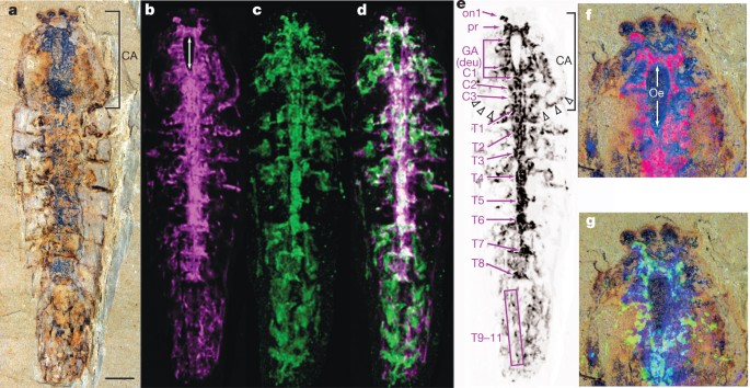
neuroanatomy in the head and trunk of a ‘great appendage’ arthropod, _Alalcomenaeus_ sp., from the Chengjiang biota, southwest China, providing the most complete neuroanatomical profile
known from a Cambrian animal. Micro-computed tomography reveals a configuration of one optic neuropil separate from a protocerebrum contiguous with four head ganglia, succeeded by eight
contiguous ganglia in an eleven-segment trunk. Arrangements of optic neuropils, the brain and ganglia correspond most closely to the nervous system of Chelicerata of all extant arthropods,
supporting the assignment of ‘great appendage’ arthropods to the chelicerate total group4,5. The position of the deutocerebral neuromere aligns with the insertion of the great appendage,
indicating its deutocerebral innervation and corroborating a homology between the ‘great appendage’ and chelicera indicated by morphological similarities4,6,7. _Alalcomenaeus_ and
_Fuxianhuia protensa_1 demonstrate that the two main configurations of the brain observed in modern arthropods, those of Chelicerata and Mandibulata, respectively8, had evolved by the early
Cambrian. Access through your institution Buy or subscribe This is a preview of subscription content, access via your institution ACCESS OPTIONS Access through your institution Subscribe to
this journal Receive 51 print issues and online access $199.00 per year only $3.90 per issue Learn more Buy this article * Purchase on SpringerLink * Instant access to full article PDF Buy
now Prices may be subject to local taxes which are calculated during checkout ADDITIONAL ACCESS OPTIONS: * Log in * Learn about institutional subscriptions * Read our FAQs * Contact customer
support SIMILAR CONTENT BEING VIEWED BY OTHERS NEUROANATOMY IN A MIDDLE CAMBRIAN MOLLISONIID AND THE ANCESTRAL NERVOUS SYSTEM ORGANIZATION OF CHELICERATES Article Open access 20 January
2022 ORDOVICIAN OPABINIID-LIKE ANIMALS AND THE ROLE OF THE PROBOSCIS IN EUARTHROPOD HEAD EVOLUTION Article Open access 15 November 2022 ORGAN SYSTEMS OF A CAMBRIAN EUARTHROPOD LARVA Article
Open access 31 July 2024 REFERENCES * Ma, X., Hou, X., Edgecombe, G. D. & Strausfeld, N. J. Complex brain and optic lobes in an early Cambrian arthropod. _Nature_ 490, 258–261 (2012)
Article ADS CAS Google Scholar * Budd, G. E. A palaeontological solution to the arthropod head problem. _Nature_ 417, 271–275 (2002) Article ADS CAS Google Scholar * Yang, J.,
Ortega-Hernández, J., Butterfield, N. J. & Zhang, X. Specialized appendages in fuxianhuiids and the head organization of early arthropods. _Nature_ 494, 468–471 (2013) Article ADS CAS
Google Scholar * Cotton, T. J. & Braddy, S. J. The phylogeny of arachnomorph arthropods and the origin of the Chelicerata. _Trans. R. Soc. Edinb. Earth Sci._ 94, 169–193 (2003)
Article Google Scholar * Stein, M., Budd, G. E., Peel, J. S. & Harper, D. A. T. _Arthroaspis_ n. gen., a common element of the Sirius Passet Lagerstätte (Cambrian, North Greenland),
sheds light on trilobite ancestry. _BMC Evol. Biol._ 13, 99 (2013) Article Google Scholar * Chen, J., Waloszek, D. & Maas, A. A new ‘great-appendage’ arthropod from the Lower Cambrian
of China and homology of chelicerate chelicerae and raptorial antero-ventral appendages. _Lethaia_ 37, 3–20 (2004) Google Scholar * Haug, J. T., Waloszek, D., Maas, A., Liu, Y. & Haug,
C. Functional morphology, ontogeny and evolution of mantis shrimp-like predators in the Cambrian. _Palaeontology_ 55, 369–399 (2012) Article Google Scholar * Strausfeld, N. J. _Arthropod
Brains: Evolution, Functional Elegance, and Historical Significance_ (Harvard Univ. Press, 2012) Google Scholar * Hou, X. & Bergström, J. Arthropods of the Lower Cambrian Chengjiang
fauna, southwest China. _Fossils and Strata_ 45, 1–116 (1997) Google Scholar * Legg, D. A., Sutton, M. D., Edgecombe, G. D. & Caron, J.-B. Cambrian bivalved arthropod reveals origins of
arthrodisation. _Proc. R. Soc. Lond. B_ 279, 4699–4704 (2012) Article Google Scholar * Edgecombe, G. D., García-Bellido, D. C. & Paterson, J. R. A new leanchoiliid megacheiran
arthropod from the lower Cambrian Emu Bay Shale, South Australia. _Acta Palaeontol. Pol._ 56, 385–400 (2011) Article Google Scholar * Haug, J. T., Briggs, D. E. G. & Haug, C.
Morphology and function in the Cambrian Burgess Shale megacheiran arthropod _Leanchoilia superlata_ and the application of a descriptive matrix. _BMC Evol. Biol._ 12, 162 (2012) Article
Google Scholar * Hou, X.-G. et al. _The Cambrian Fossils of Chengjiang, China: The Flowering of Early Animal Life_ (Blackwell, 2004) * Liu, Y., Hou, X. & Bergström, J. Chengjiang
arthropod _Leanchoilia illecebrosa_ (Hou, 1987) reconsidered. _GFF_ 129, 263–272 (2007) Article Google Scholar * Briggs, D. E. G. & Collins, D. The arthropod _Alalcomenaeus cambricus_
Simonetta, from the Middle Cambrian Burgess Shale of British Columbia. _Palaeontology_ 42, 953–977 (1999) Article Google Scholar * Brenneis, G. & Richter, S. Architecture of the
nervous system in Mystacocarida (Arthropoda, Crustacea)—an immunohistochemical study and 3D reconstruction. _J. Morphol._ 271, 169–189 (2010) PubMed Google Scholar * Damen, W. G.,
Hausdorf, M., Seyfarth, E. A. & Tautz, D. A conserved mode of head segmentation in arthropods revealed by the expression pattern of Hox genes in a spider. _Proc. Natl Acad. Sci. USA_ 95,
10665–10670 (1998) Article ADS CAS Google Scholar * García-Bellido, D. C. & Collins, D. Reassessment of the genus _Leanchoilia_ (Arthropoda, Arachnomorpha) from the Middle Cambrian
Burgess Shale, British Columbia, Canada. _Palaeontology_ 50, 693–709 (2007) Article Google Scholar * Richter, S., Stein, M., Frase, T. & Szucsich, N. U. in _Arthropod Biology and
Evolution_ (eds Minelli A., Boxshall G. & Fusco G. ) The Arthropod Head 223–240 (Springer, 2013) Google Scholar * Butterfield, N. J. _Leanchoilia_ guts and the interpretation of
three-dimensional structures in Burgess Shale-type fossils. _Paleobiology_ 28, 155–171 (2002) Article Google Scholar * Lehmann, T., Hess, M. & Melzer, R. R. Wiring a periscope -
ocelli, retinula axons, visual neuropils and the ancestrality of sea spiders. _PLoS ONE_ 7, e30474 (2012) Article ADS CAS Google Scholar * Harzsch, S. et al. Evolution of arthropod
visual systems: development of the eyes and central visual pathways in the horseshoe crab _Limulus polyphemus_ Linnaeus, 1758 (Chelicerata, Xiphosura). _Dev. Dyn._ 235, 2641–2655 (2006)
Article CAS Google Scholar * Strausfeld, N. J., Weltzien, P. & Barth, F. G. Two visual systems in one brain: neuropil serving the principal eyes of the spider _Cupiennius salei_. _J.
Comp. Neurol._ 328, 63–75 (1993) Article CAS Google Scholar * Strausfeld, N. J. & Andrew, D. R. A new view of insect-crustacean relationships. I. Inferences from neural cladistics and
comparative neuroanatomy. _Arthropod Struct. Dev._ 40, 276–288 (2011) Article Google Scholar * Zeil, J. Sexual dimorphism in the visual system of flies: the divided brain of male
Bibionidae (Diptera). _Cell Tissue Res._ 229, 591–610 (1983) Article CAS Google Scholar * Lin, C. & Strausfeld, N. J. A precocious adult visual center in the larva defines the unique
optic lobe of the split-eyed whirligig beetle _Dineutus sublineatus_. _Front. Zool._ 10, 7 (2013) Article Google Scholar * Schoenemann, B. & Clarkson, E. N. K. The eyes of
_Leanchoilia_. _Lethaia_ 45, 524–531 (2012) Article Google Scholar * Eriksson, M. E. & Terfelt, F. Exceptionally preserved Cambrian trilobite digestive system revealed in 3D by
synchrotron-radiation X-ray tomographic microscopy. _PLoS ONE_ 7, e35625 (2012) Article ADS CAS Google Scholar * Eriksson, M. E., Terfelt, F., Elofsson, R. & Marone, F. Internal
soft-tissue anatomy of Cambrian ‘Orsten’ arthropods as revealed by synchrotron X-ray tomographic microscopy. _PLoS ONE_ 7, e42582 (2012) Article ADS CAS Google Scholar Download
references ACKNOWLEDGEMENTS We thank N. Shimobayashi, H. Maeda, and T. Kogiso for arranging and performing EDXRF analyses, and D. Andrew for advice on cladistics. This work was supported by
grants from the Natural Science Foundation of China (no. 40730211), Research in Education and Science from the Government of Japan (no. 21740370), a Leverhulme Trust Research Project Grant
(F/00 696/T), by the Center for Insect Science, University of Arizona, and a grant from the Air Force Research Laboratories (FA8651-10-1-0001) to N.J.S. AUTHOR INFORMATION AUTHORS AND
AFFILIATIONS * Japan Agency for Marine-Earth Science and Technology, Yokosuka 2370061, Japan, Gengo Tanaka * Yunnan Key Laboratory for Palaeobiology, Yunnan University, Kunming 650091,
China, Xianguang Hou & Xiaoya Ma * Department of Earth Sciences, The Natural History Museum, Cromwell Road, London SW7 5BD, UK, Xiaoya Ma & Gregory D. Edgecombe * Department of
Neuroscience and Center for Insect Science, University of Arizona, Tucson, 85721, Arizona, USA Nicholas J. Strausfeld Authors * Gengo Tanaka View author publications You can also search for
this author inPubMed Google Scholar * Xianguang Hou View author publications You can also search for this author inPubMed Google Scholar * Xiaoya Ma View author publications You can also
search for this author inPubMed Google Scholar * Gregory D. Edgecombe View author publications You can also search for this author inPubMed Google Scholar * Nicholas J. Strausfeld View
author publications You can also search for this author inPubMed Google Scholar CONTRIBUTIONS The project was conceived by G.T. Fossil data were analysed by all authors. G.D.E., N.J.S. and
X.M. composed the text. CORRESPONDING AUTHORS Correspondence to Xianguang Hou or Nicholas J. Strausfeld. ETHICS DECLARATIONS COMPETING INTERESTS The authors declare no competing financial
interests. EXTENDED DATA FIGURES AND TABLES EXTENDED DATA FIGURE 1 CEPHALIC REGION OF _ALALCOMENAEUS_ SP. YKLP 11075 All in dorsal view, composites of part and counterpart (upper left).
Second left to right: CT scan (green); EDXRF Fe (red); superimposition of CT and EDXRF Fe. Lower row, left to right: EDXRF Cu (blue); superimposition of CT and EDXRF Cu; superimposition of
EDXRF Fe and EDXRF Cu; superimposition of all scans. C1, first post-GA neuropil = tritocerebrum (tri); C2, second post-GA neuropil; GA, great appendage neuropil = deutocerebrum (deu); on1,
first optic neuropil; pr, protocerebrum. EXTENDED DATA FIGURE 2 ARTHROPOD RELATIONSHIPS BASED ON NEUROANATOMICAL CHARACTERS. Strict consensus of 34 shortest cladograms based on 145
characters in Supplementary Information Table 2. SUPPLEMENTARY INFORMATION SUPPLEMENTARY INFORMATION This file contains Supplementary Tables 1-2, Phylogenetic Methods and Supplementary
References. (PDF 249 kb) SUPPLEMENTARY DATA This zipped file contains characters coded in phylogenetic analysis (in nexus format, it can be opened in freeware such as Mesquite and Nexus Data
Editor). (ZIP 3 kb) POWERPOINT SLIDES POWERPOINT SLIDE FOR FIG. 1 POWERPOINT SLIDE FOR FIG. 2 POWERPOINT SLIDE FOR FIG. 3 POWERPOINT SLIDE FOR FIG. 4 RIGHTS AND PERMISSIONS Reprints and
permissions ABOUT THIS ARTICLE CITE THIS ARTICLE Tanaka, G., Hou, X., Ma, X. _et al._ Chelicerate neural ground pattern in a Cambrian great appendage arthropod. _Nature_ 502, 364–367 (2013).
https://doi.org/10.1038/nature12520 Download citation * Received: 10 June 2013 * Accepted: 01 August 2013 * Published: 16 October 2013 * Issue Date: 17 October 2013 * DOI:
https://doi.org/10.1038/nature12520 SHARE THIS ARTICLE Anyone you share the following link with will be able to read this content: Get shareable link Sorry, a shareable link is not currently
available for this article. Copy to clipboard Provided by the Springer Nature SharedIt content-sharing initiative
