Deciphering the degradation mechanism of the lead-free all inorganic perovskite cs2sni6
- Select a language for the TTS:
- UK English Female
- UK English Male
- US English Female
- US English Male
- Australian Female
- Australian Male
- Language selected: (auto detect) - EN
Play all audios:

ABSTRACT Organic-inorganic perovskite materials are revolutionizing photovoltaics with high power conversion efficiencies, but experience significant environmental degradation and
instability. In this work, the phase stability and decomposition mechanisms of lead-free all inorganic Cs2SnI6 perovskite upon water and moisture exposure were systematically investigated
via in situ synchrotron X-ray diffraction, environmental SEM, and micro-Raman spectroscopy. A critical relative humidity (80%) is identified below which Cs2SnI6 perovskite is stable without
decomposition. Under higher humidity or aqueous environment, Cs2SnI6 perovskite decomposes into SnI4 and CsI through etch pits formation and stepwave propagation, leading to rapid crystal
dissolution. A partial reversibility of the Cs2SnI6 perovskite upon dissolution and re-precipitation with subsequent dehydration was identified, suggesting a self-healing capability of
Cs2SnI6 and thus enhanced air stability. Mechanistic understanding of the Cs2SnI6 degradation behavior can be a vital step towards developing new perovskites with enhanced environmental
stability and materials performance. SIMILAR CONTENT BEING VIEWED BY OTHERS PRESERVING A ROBUST CSPBI3 PEROVSKITE PHASE VIA PRESSURE-DIRECTED OCTAHEDRAL TILT Article Open access 19 January
2021 ADVANCES IN PHYSICOCHEMICAL CHARACTERIZATION OF LEAD-FREE HYBRID PEROVSKITE [NH3(CH2)3NH3]CUBR4 CRYSTALS Article Open access 24 May 2022 THE ROLE OF ION MIGRATION, OCTAHEDRAL TILT, AND
THE A-SITE CATION ON THE INSTABILITY OF CS1-XFAXPBI3 Article Open access 22 December 2023 INTRODUCTION Organic–inorganic perovskites have attracted significant attention from researchers in
recent years due to their impressive properties for photovoltaic applications, including high absorption coefficients, long carrier diffusion length, and solution processability,1,2,3,4
boosting the photovoltaic power conversion efficiency (PCE) over 20%.5,6,7 Despite endeavors to optimize cell design and achieve excellent device efficiencies, technical barriers to the
implementation of perovskite solar cells remain such as the toxicity of lead, and the long-term environmental instability of perovskite compositions.8 Particularly, the organic–inorganic
hybrid perovskite (CH3NH3PbI3) undergoes a rapid decomposition to PbI2 in the presence of moisture, resulting in a substantial decrease of the device performance.9,10,11,12,13 It suffers an
80% drop in PCE over a 24 h period and retains less than 5% after 6 days when the cells were stored in ambient conditions.9 In order to address the critical issues of environmental toxicity
of Pb and the associated instability, Cs2SnI6 has been explored as an alternative lead-free perovskite, which displays high degree of air stability at room temperature due to the oxidation
state of Sn4+.14,15,16 In addition to the air stability, Cs2SnI6 has a direct band gap of ~1.3 eV and high absorption coefficient (over 105 cm−1).14,15 It has been successfully adopted as a
lead-free light absorber material for solar cells and can retain over 90% in PCE for over 30 days in air.17 However, the achieved PCE of Cs2SnI6 is relatively low (~1%) in air, as compared
with CsSnI3 solar cells fabricated under tightly controlled environments,15,17 and therefore device engineering may be required to further improve the PCE. Due to the conditions expected in
the deployment of solar cells, it is vital to understand the moisture stability and the interaction behaviors of Cs2SnI6 with water or moisture. Previous work indicates that Cs2SnI6 is
stable in ambient conditions with low relative humidity (RH) (30–50% RH), and can be stored in ambient conditions for over 2 months without obvious decomposition.16 However, this material is
still susceptible to rapid decomposition upon exposure to high humidity or aqueous conditions. A mechanistic understanding of the degradation process of the lead-free all inorganic
perovskite has yet to be achieved. In this work, we report a systematic investigation of the perovskite’s degradation processes via in situ techniques with a goal of elucidating the
underlying mechanisms governing their environmental stability. Through a careful control of RH, the morphological changes were monitored using in situ environmental scanning electron
microscope (SEM). The dissolution process occurred through the formation of etch pits and subsequent generation of etching steps due to the propagation of the etch pits through the bulk
crystal. Interestingly, the well-shaped octahedra crystals recrystallized with the evaporation of water or dehydration, indicating a strong self-healing capability of Cs2SnI6. Variations in
crystallinity and phase transformations were characterized by in situ micro-Raman spectroscopy and synchrotron X-ray powder diffraction (XRD), indicating a phase decomposition of Cs2SnI6
into CsI and SnI4. The improved understanding of perovskite environmental stability and degradation maybe useful for optimization of the materials preparation and further development of new
types of perovskite with enhanced materials performance and environmental stability. RESULTS AND DISCUSSION To investigate the underlying mechanisms governing the environmental stability of
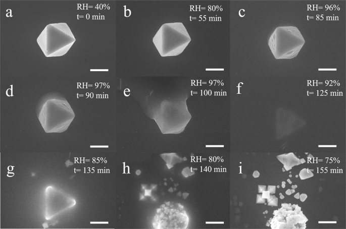
Cs2SnI6, in situ environmental SEM was performed to observe the microstructural evolution at the micro- and/or nanoscale. Figure 1 shows the in situ environmental SEM images of the
dissolution and recrystallization processes of a Cs2SnI6 crystal upon controlling the water vapor pressure (also see Supplementary Information; Supplementary Video S1). The as-prepared
Cs2SnI6 had a crystallite size ~10 µm with an octahedral shape, and was placed on an Si substrate held at 15 °C (Fig. 1a). The RH in the chamber was controlled by a leak valve. As the water
vapor pressure increased, the well-facet Cs2SnI6 crystal was maintained—even after increasing an RH to 80% in 55 min, as shown in Fig. 1b. To further confirm the phase stability in moist
air, the interaction of Cs2SnI6 with water vapor was monitored by in situ synchrotron X-ray powder diffraction under high humidity (~80%) at room temperature. For an exposure period in
excess of 7 h, in situ XRD of Cs2SnI6 powders suggests the retention of the perovskite structure, with no decomposition peaks visible, as shown in Fig. 2a. This demonstrates that Cs2SnI6 is
stable in the moist air, up to 80% RH, which is consistent with the results obtained during in situ environmental SEM and results reported previously.14,18,19 It should be noted that small
dots emerged on the crystal surface, appearing at 45% and disappearing at ~60%. These dots are believed to be Cs2SnI6 quantum dots formed during the synthesis process. As pointed out from
our previous report,16 Cs2SnI6 has significant solubility in many common organic solvents, such as ethanol, methanol, and dimethylformamide. The synthesis process in this work utilized
methanol and n-butyl acetate organic solvents. Due to the solubility of Cs2SnI6 in these organic solvents, it is highly possible that small particles re-precipitated on the crystal surface
during the drying process, as observed from the original powder (Fig. 1a). In addition, Cs2SnI6 powders prepared using ethanol in the place on n-butyl acetate displayed numerous small
particles precipitates on powders (Fig. S1). Despite the presence of these small particles across preparation techniques, all of the Cs2SnI6 powders in this study have not displayed any
impurities in the XRD characterization. Similarly, Kapil et al.20 previously reported the presences of quantum dots formed on the bulk Cs2SnI6 material. Therefore, the small dots observed in
our in situ experiment are likely quantum dots of Cs2SnI6. These small particles would easily absorb water vapor due to their high surface energy, showing high brightness. As the RH
increased to ~60%, these dots disappeared, indicating their preferential dissolution when the particle size is reduced to the nano-metered scale. Further increments of the water vapor
pressure and relatively humidity to 95% resulted in increasingly rough surfaces of the Cs2SnI6 crystals with small hillocks formed as a result of the water vapor condensation. The
dissolution process began at preferential sites instead of a uniform dissolution on the 3D crystals. The hillocks grew to a shape of small water drops and evolved into larger droplets on the
surface as the RH increased to saturated water vapor condition. Under the saturated environment, the droplets can no longer be held on the surface and instead flow on the substrate, forming
a solution around the crystal (Fig. 1c–e). Figure 1f shows the triangle-shaped residue in the dissolved solution after the crystal was held in saturated water vapor (RH > 95%) for over
30 min, suggesting a nearly complete dissolution of the original Cs2SnI6 perovskite. A reversible reaction occurs between Cs2SnI6 and water (Fig. 1f–i) with the evaporation of water
molecules to ~85% RH. The residue could act as nucleation sites from which well-shaped octahedral crystals precipitate and grow. The degradation behavior and reversible reaction of Cs2SnI6
with water were also confirmed using in situ synchrotron X-ray diffraction (XRD), as shown in Fig. 2b. Once Cs2SnI6 was exposed to water, the diffraction peaks of Cs2SnI6 perovskite
decreased rapidly. Within 10 min of water exposure, the powder was fully dissolved, and no obvious crystallinity was observed (Fig. S2). Interestingly, as the water evaporated, the
diffraction patterns which can be assigned to Cs2SnI6 reappeared, suggesting Cs2SnI6 re-precipitated from the solution (Figs 2b and S2). It is worth noting the recovered diffraction
intensity of Cs2SnI6 is lower than the original one, indicating a partial recovery process. These results indicate a critical humidity (~80% RH) exists below which the decomposition of
Cs2SnI6 will not occur and the material remains stable. The reversible reaction also suggests a strong self-healing capability of the Cs2SnI6 perovskite upon dehydration, providing strong
evidence of the enhanced environmental stability as compared with organic–inorganic hybrid perovskite.11,21,22 Detailed mechanisms of the dissolution process were further analyzed using ex
situ SEM observation, in which the formation of etch pits and dissolution surface steps were visible. In the dissolution stepwave model, the surface defects, e.g., surface steps or edge
dislocations, act as the energetically favorite sites for crystal dissolution. Figure 3a shows the fine octahedral shape of Cs2SnI6 crystals displaying a layered structure with clearly
visible growth steps, indicating the layer-by-layer growth into the bulk crystal. These surface steps and dislocations introduce strain energy into the crystal, which is energetically
favorable to the formation of etch pits.23 The dislocation strain energy can be calculated by the following equation24: $$u\left( r \right) = \frac{{\mu b^2/(8\pi ^2)}}{{r_{\mathrm h}^2 +
r^2}},$$ (1) where _µ_ is the isotropic bulk shear modulus (egs/cm3), _b_ is the size of the Burgers vector (Å), _r_h (Å) and _r_ (Å) are the size of the hollow core and the distance from
the dislocation center, respectively. Based on this equation, the center of the initial dislocation defects has a higher strain energy, and therefore the dissolution process moves away from
the center and travels across the crystal surface, generating etch pits. Further etch steps can be generated from the step far from the center of the dislocation, and the continuous movement
of these steps into the crystal will form a series of steps, based on the dissolution stepwave model.23,25 The stepwaves from each etch pit combine to lower the crystal surface area (Fig.
3b), controlling the bulk crystal dissolution rate. With the continuous dissolution, the etch pits grow in both the vertical and horizontal directions, resulting in a drop in the surface and
hollow holes in the crystals as shown in Fig. 3b, c. Figure 3d displays a schematic view of the dissolution model highlighting the formation mechanism of etch pits and the combination of
stepwaves resulting in the overall crystal dissolution. The etch pits and stepwave dissolution mechanism are more clearly visible when Cs2SnI6 was treated with methanol. Cs2SnI6 has a lower
solubility in methanol as compared to water. It is more evident that the dissolution process of Cs2SnI6 involved the formation of etch pits and the combination of stepwaves from each etch
pit eventually leads to the hollow structure of Cs2SnI6 crystals. Figure 4a shows that the triangle etch pits are visible and surrounded by relatively-flat surfaces while exposed to
methanol. The stepwaves from these etch pits combined and formed larger pits during the dissolution process (Fig. 4b). The larger pits eventually formed a large hollow pit in the crystal
with the etch pits propagating perpendicularly through the bulk crystal, as shown in Fig. 4c. A close look at the etch pit suggests that the dissolution process proceeds along the
<111> direction, forming a hexagonal dissolution pit. Figure 5 shows SEM images of various locations on the substrate after the recrystallization process, indicating the partial
reversibility of the process. In Fig. 5a, multiple octahedral crystals can be observed with a hollow structure, which are not fully dissolved under the saturated condition. These residual
crystals may act as new nucleation sites during the recrystallization process, as evidenced by the presence of small crystals on the surface (Fig. 5a). During the dissolution process, water
condensed on the substrate and formed droplets containing decomposition products. As the water evaporated, perovskite crystals re-precipitated out, forming a ring-like pattern that consisted
of small crystals, as shown in Fig. 5b. It was observed that crystals on the edge are larger than the central crystals. This may be explained by a phenomenon known as a coffee ring
effect,26 which is caused by capillary flow from the interior to the edge due to the differential evaporation rate. Dendrites and needle-like precipitates were also observed throughout the
sample, reinforcing the partial reversibility of Cs2SnI6 and the dissolution–precipitation mechanism (Fig. 5c, d). A similar partially reversible process resulting from water interaction has
also been reported in the organic–inorganic perovskite.13,22,27 Synchrotron X-ray powder diffraction was used to analyze the phase compositions of the re-precipitated materials after the
water was evaporated. As shown in Fig. 6a, the Cs2SnI6 perovskite phase is clearly visible at different locations, confirming the self-healing capability and reversible reaction with water.
In addition, SnI4 was also identified at P1, suggesting the process is not entirely reversible. Rietveld refinement of the precipitated products indicates that the phase fraction of Cs2SnI6
and SnI4 are 55% and 45%, respectively (Fig. S3). Besides, CsI could also be identified at P2 and P3. Therefore, synchrotron XRD confirms coexistence of recrystallized Cs2SnI6, and residual
decomposition products CsI and SnI4 aggregated at different regions. The CsI precipitated as large sized crystals, characterized by the diffraction spots shown in Fig. 6b. Post experiment
SEM images indicate that small particles deposited on and around the recrystallized Cs2SnI6 could be SnI4. Large dendrites were also observed similar with the in situ environmental SEM
results, which may be CsI (Fig. S4). The broad peak at 2_θ_ ~5° is attributed to the formation of Sn(OH)4, the product of the hydrolysis of SnI4. In situ Raman and optical images were also
performed to further reveal the phase decomposition and precipitation processes of Cs2SnI6 perovskite interacting with water. Figure 7a displays optical images of Cs2SnI6 powders upon the
addition of water, indicating that Cs2SnI6 can decompose rapidly upon exposure to water. Cs2SnI6 underwent a color change from black to a yellow solution and eventually a yellow slurry. The
reversibility of the dissolution–precipitation process was further supported by these color changes, with the color change from yellow to black upon drying at room temperature (Fig. 7a).
Figure 7b displays micro-Raman spectra obtained at various times after exposing Cs2SnI6 powders to water. The starting Cs2SnI6 powders had an average crystallite size around 15 µm with a
well-defined octahedral shape, as shown in Fig. 7c. For the as-prepared Cs2SnI6 powders, a sharp band at 126 cm−1 and two weak bands at 77 and 248 cm−1 were observed. The band at 126 cm−1
can be attributed to the Sn-I stretching vibration _ν_ (A1g). The low frequency band at 77 cm−1 can be ascribed to the _δ_ (F2g) mode involving I-Sn-I asymmetric bending.28 With the addition
of several water droplets, the Cs2SnI6 powders began to decompose and dissolve in water. The Raman spectrum obtained at 15 min shows broad bands at 108 and 580 cm−1, which may be attributed
to Sn(OH)429 as a result of the hydrolysis reaction of SnI4. Further dissolution results in enhanced signal from decomposition products, further evidenced by a greater number of crystals
formed at the bottom, consistent with the sharper peaks of Raman spectrum collected at 25 min. As water evaporated, Cs2SnI6 crystals re-precipitated from the solution as shown in Fig. 7b. In
addition, the strong peak at 145 cm−1 emerged due to the presence of SnI4, consistent with the findings from synchrotron XRD diffraction (Fig. 6a). The strong variation in color between the
two compositions, the black crystal is Cs2SnI6 and the orange particles are SnI4, enabling their identification via optical microscopy in Fig. 7f. Optical images shown in Fig. 8a display
the decomposition of Cs2SnI6 in water, as evidenced by the white gel-like precipitates which are the result of a hydrolysis reaction forming tin (IV) hydroxide. Changes in the pH value
suggest a strong acid solution arising from the formation of hydriodic acid (Fig. 8c) associated with the hydrolysis reaction (reaction 3). A similar phenomena was observed in a prior tin
(IV) iodide hydrolysis experiment.30 After 12 h, the transparent solution turned yellow, indicating the presence of molecular iodine in the solution (Fig. 8b). The formation of molecular
iodine may be attributed to the decomposition product of hydrogen iodide. Based on the systematic in situ measurements of phases, morphology evolution and solution chemistry, the dissolution
and recrystallization mechanism of the lead-free all inorganic Cs2SnI6 perovskite upon water interaction can be expressed as follows: $${\mathrm{Cs}}_2{\mathrm{SnI}}_6 \leftrightarrow
{\mathrm{SnI}}_4 + 2{\mathrm{CsI}},$$ (2) $${\mathrm{SnI}}_4 + 4{\mathrm{H}}_2{\mathrm{O}} \leftrightarrow {\mathrm{Sn}}({\mathrm{OH}})_4 + 4{\mathrm{HI}},$$ (3) Finally, the degradation
behavior of Cs2SnI6 was compared to the organic–inorganic perovskite (MAPbI3 or CH3NH3PbI3) to further improve the general understanding of perovskite degradation mechanisms. For MAPbI3
perovskite, during the reversible hydration process, water molecules are incorporated into the crystal structure, forming the monohydrate (CH3NH3PbI3·H2O).22,31,32 With prolonged water vapor
exposure, it forms the dihydrate, (CH3NH3)4PbI6·2H2O), leading to an irreversible degradation of MAPbI3 perovskite.22,32 The water molecule intercalation into the crystal structure induces
the distortion of the 3D MAPbI3 perovskite structure and rearrangement, resulting in a separation of the [PbI6]4− octahedra to form 1D double-chains MAPbI3 perovskite species or 0D
frameworks.22 However, in the Cs2SnI6 perovskite no hydrated phases have been observed. This may be explained by the higher hydration energy of the Cs+ cation (−67.7 kcal/mol),33 as compared
to the CH3NH3+ cation (−75 kcal/mol).34 In addition, due to the high ionic mobility of Cs+ cation in water,35 Cs+ cations may diffuse out more rapidly upon exposure to water resulting in
the dissociation of the Cs2SnI6 perovskite crystal structure, rather than forming the hydrates. The Sn4+ is stable in the ambient condition, but experiences the hydrolysis in the water.
Therefore, the enhanced moisture stability of Cs2SnI6 as compared with that of the organic–inorganic perovskite may be a result of synergistic effects from both Cs+ and Sn4+, and more
investigations are needed to determine the relative contributions of Cs, Sn, and I to the stability. In summary, this work investigated the degradation process of Cs2SnI6 perovskite under
controlled RH environment or aqueous conditions via in situ measurements, such as in situ environmental SEM, in situ synchrotron XRD, and Raman spectroscopy. The results demonstrate that
Cs2SnI6 is stable below a critical relatively humidity of 80%, but experiences degradation when exposed to moisture in excess of 80% RH or aqueous conditions. The degradation process of
Cs2SnI6 was attributed to etch pit formation on the surface of crystals, resulting in the dissolution of the entire crystal as explained by the stepwave model. Through in situ synchrotron
XRD and micro-Raman spectroscopy, it was determined that Cs2SnI6 decomposes into CsI and SnI4 in water as evidenced by the formation of the hydrolysis product Sn(OH)4 in the solution. These
results may elucidate the fundamental decomposition pathways of Cs2SnI6 perovskite governing the stability of halide perovskite. METHODS MATERIALS Unless stated otherwise, all materials and
solvents are used as-purchased without further purification. Cesium iodide (CsI, 99.9%), n-butyl acetate (99% min), and hydroiodic acid (HI, 55–58%) were purchased from Alfa Aesar. Tin
powder (Sn, ≥99%) and Iodine (I2, ≥99.99%) were from Sigma Aldrich. CS2SNI6 PREPARATION The method follows as previous reported.16 Typically, CsI (1 mmol) was dissolved in 10 mL methanol and
heated to ~60 °C in a water bath. In a separated 25 mL beaker, SnI4 (0.5 mmol) was dissolved in 4 mL n-butyl acetate with addition of 2 mL hydroiodic acid. Addition of the acid SnI4
solution to the warm CsI solution under vigorous stirring led to spontaneous precipitation of a fine black powder. The mixture was stirred for a further 30 min to ensure completion of the
reaction in the 60 °C water bath. Then the solid was washed by n-butyl acetate twice via centrifuging and dried in an oven at 80 °C overnight at ambient conditions. IN SITU ENVIRONMENTAL SEM
TEST Environmental SEM (FEI, Versa 3D Dualbeam) was used for the in situ analysis of the dissolution and recrystallization process of Cs2SnI6 via controlling RH. Water vapor was introduced
into the chamber through a leak valve. Cs2SnI6 crystals were placed on an Si substrate controlled at 15 °C. For the in situ experiment, first the water vapor pressure was set to be around
600 Pa to achieve an RH of ~35%. Then, the pressure was increased at a constant rate (20 Pa/min) to a value 1000 Pa (RH = 58.5%), followed by a rate at 10 Pa/min until reaching 1660 Pa. The
pressure was then held for 20 min to allow crystal dissolution. After that, the water vapor pressure was reduced at a constant rate of 10 Pa/min to a value ~715 Pa (RH = 41%). During the
experiment, images were captured manually with a speed at every minute or every 30 s as appropriate for the reaction kinetics. Without further notification, each image is of the same
magnification, taken at the same location. IN SITU SYNCHROTRON POWDER DIFFRACTION DATA COLLECTION In situ synchrotron powder XRD was performed at the Advanced Photon Source on beamline
17-BM-B at Argonne National Laboratory using an Si (111) monochromator (_λ_ = 0.39433 Å). The experiment was carried out by loading approximately 5 mg of powder sample in a Kapton tubing of
1.1 mm diameter. After the diffraction patterns of dry powders were taken, ~250 µL water was added on the powder directly to monitor the degradation process of Cs2SnI6 in aqueous condition.
It should be noted that after water addition it takes about 5 min before the first pattern obtained (i.e. time between seventh and eighth diffraction pattern), due to the time for restarting
the system. The diffraction pattern was recorded by an amorphous silicon-based area detector from PerkinElmer (2048 × 2048, 200 µm pixels). All data were collected at room temperature. To
monitor the reaction at real time, the diffraction patterns were continuously collected with 60 s per frame. The obtained patterns were analyzed using the Rietveld refinement functions
implemented in GSAS II.36,37 In situ micro-Raman spectra were measured on a Renishaw Micro-Raman spectrometer equipped with an Ar+ ion laser (_λ_ = 514.5 nm) at room temperature. The spectra
were collected at every 5 min interval to monitor the interaction between Cs2SnI6 and water. A typical spectrum was acquired at an exposure time of 30 s and two accumulations under a power
of 20 mW. To investigate the Cs2SnI6 morphology changes before/after the water reaction, SEM was carried out on a Carl Zeiss Supra 55 field emission scanning electron microscope. DATA
AVAILABILITY The data that support the findings of this study are available from the corresponding author upon reasonable request. REFERENCES * Wehrenfennig, C., Eperon, G. E., Johnston, M.
B., Snaith, H. J. & Herz, L. M. High charge carrier mobilities and lifetimes in organolead trihalide perovskites. _Adv. Mater._ 26, 1584–1589 (2014). Article CAS Google Scholar *
Xing, G. et al. Long-range balanced electron-and hole-transport lengths in organic-inorganic CH3NH3PbI3. _Science_ 342, 344–347 (2013). Article CAS Google Scholar * Jeon, N. J. et al.
Solvent engineering for high-performance inorganic–organic hybrid perovskite solar cells. _Nat. Mater._ 13, 897–903 (2014). Article CAS Google Scholar * Dong, Q. et al. Electron-hole
diffusion lengths >175 μm in solution-grown CH3NH3PbI3 single crystals. _Science_ 347, 967–970 (2015). Article CAS Google Scholar * Jiang, Q. et al. Planar-structure perovskite solar
cells with efficiency beyond 21%. _Adv. Mater._ 29, 1703852 (2017). Article Google Scholar * Yang, W. S. et al. High-performance photovoltaic perovskite layers fabricated through
intramolecular exchange. _Science_ 348, 1234–1237 (2015). Article CAS Google Scholar * Saliba, M. et al. Incorporation of rubidium cations into perovskite solar cells improves
photovoltaic performance. _Science_ 354, 206–209 (2016). Article CAS Google Scholar * Hodes, G. & Cahen, D. Photovoltaics: perovskite cells roll forward. _Nat. Photonics_ 8, 87–88
(2014). Article CAS Google Scholar * Zhou, H. et al. Interface engineering of highly efficient perovskite solar cells. _Science_ 345, 542–546 (2014). Article CAS Google Scholar * Han,
Y. et al. Degradation observations of encapsulated planar CH3NH3PbI3 perovskite solar cells at high temperatures and humidity. _J. Mater. Chem. A_ 3, 8139–8147 (2015). Article CAS Google
Scholar * Yang, J., Siempelkamp, B. D., Liu, D. & Kelly, T. L. Investigation of CH3NH3PbI3 degradation rates and mechanisms in controlled humidity environments using in situ techniques.
_ACS Nano_ 9, 1955–1963 (2015). Article CAS Google Scholar * Christians, J. A., Miranda Herrera, P. A. & Kamat, P. V. Transformation of the excited state and photovoltaic efficiency
of CH3NH3PbI3 perovskite upon controlled exposure to humidified air. _J. Am. Chem. Soc._ 137, 1530–1538 (2015). Article CAS Google Scholar * Zhao, J. et al. Investigation of the
hydrolysis of perovskite organometallic halide CH3NH3PbI3 in humidity environment. _Sci. Rep._ 6, 21976 (2016). Article CAS Google Scholar * Lee, B. et al. Air-stable molecular
semiconducting iodosalts for solar cell applications: Cs2SnI6 as a hole conductor. _J. Am. Chem. Soc._ 136, 15379–15385 (2014). Article CAS Google Scholar * Qiu, X. et al. From unstable
CsSnI3 to air-stable Cs2SnI6: a lead-free perovskite solar cell light absorber with bandgap of 1.48 eV and high absorption coefficient. _Sol. Energy Mater. Sol. Cells_ 159, 227–234 (2017).
Article CAS Google Scholar * Zhu, W. et al. Tunable optical properties and stability of lead free all inorganic perovskites (Cs2SnIxCl6-x). _J. Mater. Chem. A_ 6, 2577–2584 (2018).
Article CAS Google Scholar * Lee, B., Krenselewski, A., Baik, S. I., Seidman, D. N. & Chang, R. P. Solution processing of air-stable molecular semiconducting iodosalts, Cs2SnI6−xBrx,
for potential solar cell applications. _Sustain. Energy Fuels_ 1, 710–724 (2017). Article CAS Google Scholar * Jiang, Y., Zhang, H., Qiu, X. & Cao, B. The air and thermal stabilities
of lead-free perovskite variant Cs2SnI6 powder. _Mater. Lett._ 199, 50–52 (2017). Article CAS Google Scholar * Guo, F. et al. A two-step dry process for Cs2SnI6 perovskite thin film.
_Mater. Res. Lett._ 5, 540–546 (2017). Article CAS Google Scholar * Kapil, G. et al. Investigation of interfacial charge transfer in solution processed Cs2SnI6 thin films. _J. Phys. Chem.
C_ 121, 13092–13100 (2017). Article CAS Google Scholar * Wang, Q. et al. Scaling behavior of moisture-induced grain degradation in polycrystalline hybrid perovskite thin films. _Energy
Environ. Sci._ 10, 516–522 (2017). Article CAS Google Scholar * Leguy, A. l. M. et al. Reversible hydration of CH3NH3PbI3 in films, single crystals, and solar cells. _Chem. Mater._ 27,
3397–3407 (2015). Article CAS Google Scholar * Lasaga, A. C. & Lü̈ttge, A. A model for crystal dissolution. _Eur. J. Mineral._ 15, 603–615 (2003). Article CAS Google Scholar *
Heinisch, H., Sines, G., Goodman, J. & Kirby, S. Elastic stresses and self-energies of dislocations of arbitrary orientation in anisotropic media: olivine, orthopyroxene, calcite, and
quartz. _J. Geophys. Res._ 80, 1885–1896 (1975). Article Google Scholar * Lasaga, A. C. & Luttge, A. Variation of crystal dissolution rate based on a dissolution stepwave model.
_Science_ 291, 2400–2404 (2001). Article CAS Google Scholar * Deegan, R. D. et al. Capillary flow as the cause of ring stains from dried liquid drops. _Nature_ 389, 827–829 (1997).
Article CAS Google Scholar * Song, Z. et al. Perovskite solar cell stability in humid air: partially reversible phase transitions in the PbI2-CH3NH3I-H2O system. _Adv. Energy Mater_ 6,
1600846, 1–7 (2016). Google Scholar * Kaltzoglou, A. et al. Optical-vibrational properties of the Cs2SnX6 (X = Cl, Br, I) defect perovskites and hole-transport efficiency in dye-sensitized
solar cells. _J. Phys. Chem. C._ 120, 11777–11785 (2016). Article CAS Google Scholar * Huang, B. X., Tornatore, P. & Li, Y.-S. IR and Raman spectroelectrochemical studies of corrosion
films on tin. _Electrochim. Acta_ 46, 671–679 (2001). Article Google Scholar * Hickling, G. G. Gravimetric analysis: the synthesis of tin iodide. _J. Chem. Educ._ 67, 702 (1990). Article
CAS Google Scholar * Hao, F., Stoumpos, C. C., Liu, Z., Chang, R. P. & Kanatzidis, M. G. Controllable perovskite crystallization at a gas–solid interface for hole conductor-free
solar cells with steady power conversion efficiency over 10%. _J. Am. Chem. Soc._ 136, 16411–16419 (2014). Article CAS Google Scholar * Schlipf, J. et al. In situ monitoring the uptake of
moisture into hybrid perovskite thin films. _J. Phys. Chem. Lett._ 9, 2015–2021 (2018). Article CAS Google Scholar * Aqvist, J. Ion-water interaction potentials derived from free energy
perturbation simulations. _J. Phys. Chem._ 94, 8021–8024 (1990). Article Google Scholar * Aue, D. H., Webb, H. M. & Bowers, M. T. A thermodynamic analysis of solvation effects on the
basicities of alkylamines. An electrostatic analysis of substituent effects. _J. Am. Chem. Soc._ 98, 318–329 (1976). Article CAS Google Scholar * Lee, S. H. & Rasaiah, J. C. Molecular
dynamics simulation of ion mobility. 2. Alkali metal and halide ions using the SPC/E model for water at 25 C. _J. Phys. Chem._ 100, 1420–1425 (1996). Article CAS Google Scholar *
Albinati, A. & Willis, B. The Rietveld method in neutron and X-ray powder diffraction. _J. Appl. Crystallogr._ 15, 361–374 (1982). Article CAS Google Scholar * Toby, B. H. & Von
Dreele, R. B. GSAS-II: the genesis of a modern open-source all purpose crystallography software package. _J. Appl. Crystallogr._ 46, 544–549 (2013). Article CAS Google Scholar Download
references ACKNOWLEDGEMENTS The synthesis of Cs2SnI6 perovskite and dissolution mechanistic understandings were supported as part of the Center for Performance and Design of Nuclear Waste
Forms and Containers, an Energy Frontier Research Center funded by the U.S. Department of Energy, Office of Science, Basic Energy Sciences under Award # DE-SC0016584. In situ Synchrotron
X-ray diffraction was performed using Beamline 17-BM of the Advanced Photon Source (APS), a US DOE Office of Science User Facility operated for the DOE Office of Science by Argonne National
Laboratory under contract no. DE-AC02-06CH11357. AUTHOR INFORMATION AUTHORS AND AFFILIATIONS * Department of Mechanical, Aerospace and Nuclear Engineering, Rensselaer Polytechnic Institute,
Troy, NY, 12180, USA Weiguang Zhu, Guoqing Xin, Spencer M. Scott, Tiankai Yao, Bowen Gong, Yachun Wang, Mingxin Li & Jie Lian * X-ray Science Division, Advanced Photon Source, Argonne
National Laboratory, Lemont, IL, 60439, USA Wenqian Xu Authors * Weiguang Zhu View author publications You can also search for this author inPubMed Google Scholar * Guoqing Xin View author
publications You can also search for this author inPubMed Google Scholar * Spencer M. Scott View author publications You can also search for this author inPubMed Google Scholar * Wenqian Xu
View author publications You can also search for this author inPubMed Google Scholar * Tiankai Yao View author publications You can also search for this author inPubMed Google Scholar *
Bowen Gong View author publications You can also search for this author inPubMed Google Scholar * Yachun Wang View author publications You can also search for this author inPubMed Google
Scholar * Mingxin Li View author publications You can also search for this author inPubMed Google Scholar * Jie Lian View author publications You can also search for this author inPubMed
Google Scholar CONTRIBUTIONS The scope of the experiment was designed by W.Z. and J.L. W.Z. carried out the material synthesis, micro-Raman spectroscopy. G.X. and S.M.S. helped with the
sample preparation. W.Z., W.X., T.Y., and B.G. conducted in the in situ synchrotron X-ray diffraction. W.Z. and T.Y. performed in situ environmental SEM and morphology characterization Y.W.
was involved in Raman data analysis. M.L. helped draw the schematic graph. W.Z. and J.L. wrote the manuscript. All the authors helped on the paper editing. CORRESPONDING AUTHOR
Correspondence to Jie Lian. ETHICS DECLARATIONS COMPETING INTERESTS The authors declare no competing interests. ADDITIONAL INFORMATION PUBLISHER’S NOTE: Springer Nature remains neutral with
regard to jurisdictional claims in published maps and institutional affiliations. SUPPLEMENTARY INFORMATION SUPPLEMENTARY INFORMATION. SUPPLEMENTARY VIDEO S1. RIGHTS AND PERMISSIONS OPEN
ACCESS This article is licensed under a Creative Commons Attribution 4.0 International License, which permits use, sharing, adaptation, distribution and reproduction in any medium or format,
as long as you give appropriate credit to the original author(s) and the source, provide a link to the Creative Commons license, and indicate if changes were made. The images or other third
party material in this article are included in the article’s Creative Commons license, unless indicated otherwise in a credit line to the material. If material is not included in the
article’s Creative Commons license and your intended use is not permitted by statutory regulation or exceeds the permitted use, you will need to obtain permission directly from the copyright
holder. To view a copy of this license, visit http://creativecommons.org/licenses/by/4.0/. Reprints and permissions ABOUT THIS ARTICLE CITE THIS ARTICLE Zhu, W., Xin, G., Scott, S.M. _et
al._ Deciphering the degradation mechanism of the lead-free all inorganic perovskite Cs2SnI6. _npj Mater Degrad_ 3, 7 (2019). https://doi.org/10.1038/s41529-019-0068-3 Download citation *
Received: 16 August 2018 * Accepted: 10 January 2019 * Published: 15 February 2019 * DOI: https://doi.org/10.1038/s41529-019-0068-3 SHARE THIS ARTICLE Anyone you share the following link
with will be able to read this content: Get shareable link Sorry, a shareable link is not currently available for this article. Copy to clipboard Provided by the Springer Nature SharedIt
content-sharing initiative